Korean J Obstet Gynecol.
2010 May;53(5):443-448. 10.5468/kjog.2010.53.5.443.
Twin pregnancy with diploid partial hydatidiform mole and coexisting fetuses following in vitro fertilization and embryo transfer: A case report
- Affiliations
-
- 1Department of Obstetrics and Gynecology, Chonbuk National University Medical School, Jeonju, Korea. yjjeong@jbnu.ac.kr
- 2Research Institute of Clinical Medicine, Chonbuk National University Medical School, Jeonju, Korea.
- KMID: 2273877
- DOI: http://doi.org/10.5468/kjog.2010.53.5.443
Abstract
- A 24-year-old woman, gravida 1, para 0, was referred to our hospital at 14 weeks of gestation due to suspected twin pregnancy with hydatidiform mole and coexisting fetuses. The present pregnancy was achieved following in vitro fertilization and embryo transfer (IVF-ET). Ultrasound examinations at 14 weeks 3 days of gestation revealed a live fetus appropriate for assigned gestational age with a normal-looking placenta and a dead fetus with an additional echogenic mass resembling molar placenta. The patient was planned to take amniocentesis for chromosomal analysis. However, regular uterine contraction was developed and spontaneous expulsion was occurred at 14 weeks 4 days of gestation. Chromosomal analysis of twin pregnancy using normal and molar placental tissues revealed normal karyotype with 46,XY, 46,XX, respectively. Follow-up showed no progression to persistent gestational trophoblastic disease. We present a twin pregnancy with diploid partial hydatidiform mole and coexisting fetuses that occurred following IVF-ET, which was aborted spontaneously.
Keyword
MeSH Terms
Figure
-
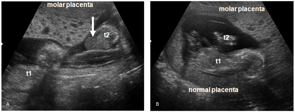
Fig. 1 Transabdominal ultrasonographic findings at 14 weeks 3 days of gestation. (A) A uterus contains diffuse multiple hypoechoic cysts. An omphalocele (arrow) is seen. (B) A coexisted live fetus with normal placenta is seen in the uterus. t1:twin 1, t2: twin 2.

Fig. 2 Postmortem photograph. Ruptured omphalocele (arrow) is seen. t1:twin 1, t2: twin 2.

Fig. 3 Pathologic findings. (A) Gross picture of the molar placenta (arrow) showing a diffuse cluster of molar vesicles. (B) Microscopic picture of the molar placenta showing an admixture of normal villi (arrow head) and molar villi displaying scalloped outlines (H&E stain, ×100).
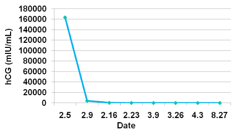
Fig. 4 The course of β-hCG levels after spontaneous abortion.
Reference
-
1. Sebire NJ, Fisher RA, Rees HC. Histopathological diagnosis of partial and complete hydatidiform mole in the first trimester of pregnancy. Pediatr Dev Pathol. 2003. 6:69–77.2. Vassilakos P, Riotton G, Kajii T. Hydatidiform mole: two entities. A morphologic and cytogenetic study with some clinical consideration. Am J Obstet Gynecol. 1977. 127:167–170.3. Szulman AE, Surti U. The syndromes of hydatidiform mole. I. Cytogenetic and morphologic correlations. Am J Obstet Gynecol. 1978. 131:665–671.4. Fluker MR, Yuzpe AA. Partial hydatidiform mole following transfer of a cryopreserved-thawed blastocyst. Fertil Steril. 2000. 74:828–829.5. Moon IK, Seo HS, Shin HM, Chun GH, Ahn YJ, Song HY, et al. A case of advanced twin pregnancy with partial hydatidiform mole. Korean J Obstet Gynecol. 1996. 39:201–205.6. Chu W, Chapman J, Persons DL, Fan F. Twin pregnancy with partial hydatidiform mole and coexistent fetus. Arch Pathol Lab Med. 2004. 128:1305–1306.7. Lee YK, Sung HS, Hong JS, Shim SS, Park JS, Jun JK, et al. A case of partial hydatidiform mole with coexisting twin pregnancy following in vtro fertilization and embryo transfer. Korean J Obstet Gynecol. 2004. 47:1586–1590.8. Ingec M, Borekci B, Altas S, Kadanali S. Twin pregnancy with partial hydatidiform mole and coexistent normal fetus. J Obstet Gynaecol. 2006. 26:379–380.9. Steller MA, Genest DR, Bernstein MR, Lage JM, Goldstein DP, Berkowitz RS. Clinical features of multiple conception with partial or complete molar pregnancy and coexisting fetuses. J Reprod Med. 1994. 39:147–154.10. Sebire NJ, Rees H, Paradinas F, Seckl M, Newlands E. The diagnostic implications of routine ultrasound examination in histologically confirmed early molar pregnancies. Ultrasound Obstet Gynecol. 2001. 18:662–665.11. Fowler DJ, Lindsay I, Seckl MJ, Sebire NJ. Routine pre-evacuation ultrasound diagnosis of hydatidiform mole: experience of more than 1000 cases from a regional referral center. Ultrasound Obstet Gynecol. 2006. 27:56–60.12. Wee L, Jauniaux E. Prenatal diagnosis and management of twin pregnancies complicated by a co-existing molar pregnancy. Prenat Diagn. 2005. 25:772–776.13. Feinberg RF, Lockwood CJ, Salafia C, Hobbins JC. Sonographic diagnosis of a pregnancy with a diffuse hydatidiform mole and coexistent 46,XX fetus: a case report. Obstet Gynecol. 1988. 72:485–488.14. Vejerslev LO. Clinical management and diagnostic possibilities in hydatidiform mole with coexistent fetus. Obstet Gynecol Surv. 1991. 46:577–588.15. Berkowitz RS, Goldstein DP. Berek JS, editor. Gestational trophoblastic disease. Berek & Novak's Gynecology. 2007. 14th ed. Philadelphia: Lippincott Williams & Wilkins;1588.16. Sebire NJ, Foskett M, Paradinas FJ, Fisher RA, Francis RJ, Short D, et al. Outcome of twin pregnancies with complete hydatidiform mole and healthy co-twin. Lancet. 2002. 359:2165–2166.17. Jauniaux E. Partial moles: from postnatal to prenatal diagnosis. Placenta. 1999. 20:379–388.18. Lage JM, Berkowitz RS, Rice LW, Goldstein DP, Bernstein MR, Weinberg DS. Flow cytometric analysis of DNA content in partial hydatidiform moles with persistent gestational trophoblastic tumor. Obstet Gynecol. 1991. 77:111–115.19. Davis JR, Kerrigan DP, Way DL, Weiner SA. Partial hydatidiform moles: deoxyribonucleic acid content and course. Am J Obstet Gynecol. 1987. 157:969–973.20. Teng NN, Ballon SC. Partial hydatidiform mole with diploid karyotype: report of three cases. Am J Obstet Gynecol. 1984. 150:961–964.
- Full Text Links
-
- Actions
-
Cited
- CITED
-
- Close
- Share
-
- Similar articles
-
- A Case of Partial Hydatidiform Mole with Coexisting Twin Pregnancy Following In Vitro Fertilization and Embryo Transfer
- A Case of Complete Hydatidiform Mole in a triplet pregnancy following In Vitro Fertilization and Embryo Transfer
- A Case of Twin Pregnancy with Complete Hydatidiform Mole and Coexisting Fetus Following IVF-ET
- A Case of Twin Pregnancy Consisting of Complete Hydatidiform Mole and a Coexisting Fetus Following IUI
- Complete Hydatidiform Mole Coexisting Two Live Fetuses in Triplet Pregnancy
