Korean J Obstet Gynecol.
2010 Mar;53(3):219-226. 10.5468/kjog.2010.53.3.219.
The predictive value of abnormal ultrasonographic finding for fetal trisomy in the second trimester
- Affiliations
-
- 1Department of Obstetrics and Gynecology, Chonbuk National University Medical School, Jeonju, Korea. jh.lee@jbnu.ac.kr
- KMID: 2273847
- DOI: http://doi.org/10.5468/kjog.2010.53.3.219
Abstract
OBJECTIVE
The purpose of this study is to evaluate obstetrical characteristics related to fetal trisomies and to survey the predictive value of abnormal second trimester ultrasonographic findings for fetal trisomies.
METHODS
We reviewed the medical records of 3,023 patients who had fetal karyotyping performed between May 1989 and May 2005, and then retrospectively examined 71 cases of trisomies diagnosed prenatally. All patients were classified into three groups according to indications of fetal karyotyping such as positive triple test result, maternal age older than 35 at delivery, and abnormal ultrasonographic findings and we compared the obstetrical features and positive predictive value of each indication.
RESULTS
Thirty two cases (39%) of total trisomies had abnormal ultrasonographic findings. Abnormal ultrasonographic findings were significantly more common (16 cases, 76%, P=0.001) in fetuses with trisomy 18 compared to the other trisomies. Structural anomalies in fetuses with trisomy were usually detected in late second trimester. The positive predictive value of abnormal ultrasonographic findings was 3.0% (elderly woman; 1.4%, positive triple test; 1.7%) in trisomy 21 and 6.3% (elderly woman; 1.6%, positive triple test; 1.8%) in both trisomy 21 and 18.
CONCLUSION
The positive predictive value of abnormal ultrasonographic findings for diagnosis of fetal trisomy is higher than the other indications for fetal karyotyping. In addition, screening of trisomy 18 with an ultrasonography may be still more important because the majority of fetuses with trisomy 18 show various congenital anomalies.
MeSH Terms
Figure
-
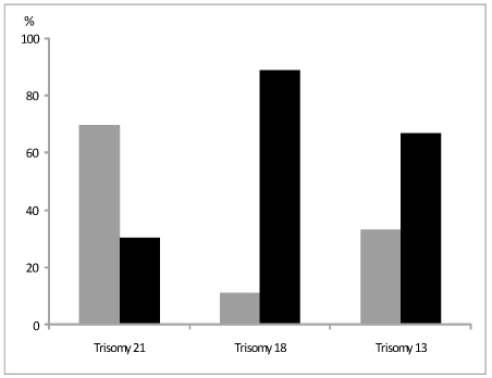
Figure 1 Percentage of ultrasonographic findings (▒ normal, ▪ abnormal) in various trisomies.
Reference
-
1. Hassold TJ, Schwartz S. Fauci AS, Braunwald E, Kasper DL, editors. Chromosome disorders. Harrison's principles of internal medicine. 2008. 17th ed. New York: McGraw-Hill.2. Hook EB, Cross PK, Schreinemachers DM. Chromosomal abnormality rates of amniocentesis and in live-born infants. JAMA. 1983. 249:2034–2038.3. Merkatz IR, Nitowsky HM, Macri JN, Johnson WE. An association between low maternal serum α-fetoprotein and fetal chromosomal abnormalities. Am J Obstet Gynecol. 1984. 148:886–894.
Article4. Malone FD, Canick JA, Ball RH, Nyberg DA, Comstock CH, Bukowski R, et al. First-trimester or second-trimester screening, or both, for Down's syndrome. N Engl J Med. 2005. 353:2001–2011.
Article5. Nicolaides KH. Nuchal translucency and other first-trimester sonographic markers of chromosomal abnormalities. Review article. Am J Obstet Gynecol. 2004. 191:45–67.7. Nyberg DA, Souter VL. Sonographic markers of fetal trisomies: second trimester. J Ultrasound Med. 2001. 20:655–674.
Article8. Little BB, Ramin SM, Cambridge BS, Schneider NR, Cohen DS, Snell LM, et al. Risk of chromosomal abnormalities, with emphasis on live-born offspring of young mothers. Am J Hum Genet. 1995. 57:1178–1185.9. American College of Obstetricians and Gynecologists (ACOG). Screening for fetal chromosomal abnormalities. 2007. 01. Washington (DC): American College of Obstetricians and Gynecologists (ACOG);11. (ACOG practice bulletin; no. 77).10. Phillips OP, Elias S, Shulman LP, Andersen RN, Morgan CD, Simpson JL. Maternal serum screening for fetal Down syndrome in women less than 35 years of age using α-fetoprotein, hCG, and unconjugated estriol: a prospective 2-year study. Obstet Gynecol. 1992. 80(3 Pt 1):353–358.11. Malone FD, Wald NJ, Canick JA, Ball RH, Nyberg DA, Comstock CH, et al. First and second trimester evaluation of risk (FASTER) trial: Principal results of the NICHD multicenter Down syndrome screening study. Am J Obstet Gynecol. 2003. 189:6 suppl 1. S56.12. Cuckle H, Benn P, Wright D. Down syndrome screening in the first and/or second trimester: Model predicted performance using meta-analysis parameters. Semin Perinatol. 2005. 29:252–257.
Article13. Vintzileos AM, Egan JF. Adjusting the risk for trisomy 21 on the basis of second-trimester ultrasonography. Am J Obstet Gynecol. 1995. 172:837–844.
Article14. Benacerraf BR, Barss VA, Laboda LA. A sonographic sign for the detection in the second trimester of the fetus with Down's syndrome. Am J Obstet Gynecol. 1985. 151:1078–1079.
Article15. Yeo L, Guzman ER, Day-Salvatore D, Walters C, Chavez D, Vintzileos AM. Prenatal detection of fetal trisomy 18 through abnormal sonographic features. J Ultrasound Med. 2003. 22:581–590. quiz 591-2.
Article16. Sohl BD, Scioscia AL, Budorick NE, Moore TR. Utility of minor ultrasonographic markers in the prediction of abnormal fetal karyotype at a prenatal diagnostic center. Am J Obstet Gynecol. 1999. 181:898–903.
Article17. Park SY, Kim JW, Kim YM, Kim JM, Lee MH, Lee BY, et al. Frequencies of fetal chromosomal abnormalities at prenatal diagnosis: 10 years experiencies in a single institution. J Korean Med Sci. 2001. 16:290–293.18. Palomaki GE, Haddow JE, Knight GJ, Wald NJ, Kennard A, Canick JA, et al. Risk-based prenatal screening for trisomy 18 using alpha-fetoprotein, unconjugated oestriol and human chorionic gonadotropin. Prenat Diagn. 1995. 15:713–723.
Article19. Bundy AL, Saltzman DH, Pober B, Fine C, Emerson D, Doubilet PM. Antenatal sonographic findings in trisomy 18. J Ultrasound Med. 1986. 5:361–364.
Article20. Benacerraf BR, Miller WA, Frigoletto FD Jr. Sonographic detection of fetuses with trisomies 13 and 18: accuracy and limitations. Am J Obstet Gynecol. 1988. 158:404–409.
Article21. Hook EB, Woodbury DF, Albright SG. Rates of trisomy 18 in livebirths, stillbirths, and at amniocentesis. Birth Defects Orig Artic Ser. 1979. 15:81–93.24. Yagel S, Anteby EY, Hochner-Celnikier D, Ariel I, Chaap T, Ben Neriah Z. The role of midtrimester targeted fetal organ screening combined with the "triple test" and maternal age in the diagnosis of trisomy 21: A retrospective study. Am J Obtet Gynecol. 1998. 178:40–44.
Article
- Full Text Links
-
- Actions
-
Cited
- CITED
-
- Close
- Share
-
- Similar articles
-
- Cytogenetic and clinical analysis of midtrimester amniocentesis
- A case of the Patau syndrome diagnosed in second trimester
- A Case of Prenatally Detected Trisomy 9 Associated with Dandy Walker Syndrome
- Recurrent Partial Trisomy 1q in Maternal Balanced Translocation t(1;11)(q32;q23)
- ACase of the Patau Syndrome Diagnosed in Second Trimester
