Branching Patterns of Medial and Inferior Calcaneal Nerves Around the Tarsal Tunnel
- Affiliations
-
- 1Department of Physical Medicine and Rehabilitation, Korea University College of Medicine, Seoul, Korea. rmkdh@korea.ac.kr
- 2Department of Anatomy, Korea University College of Medicine, Seoul, Korea.
- KMID: 2273019
- DOI: http://doi.org/10.5535/arm.2015.39.1.52
Abstract
OBJECTIVE
To demonstrate the bifurcation pattern of the tibial nerve and its branches.
METHODS
Eleven legs of seven fresh cadavers were dissected. The reference line for the bifurcation point of tibial nerve branches was an imaginary horizontal line passing the tip of the medial malleolus. The distances between the reference line and the bifurcation points were measured. The bifurcation branching patterns were categorized as type I, the pattern in which the medial calcaneal nerve (MCN) branched most proximally; type II, the pattern in which the three branches occurred at the same point; and type III, in which MCN branched most distally.
RESULTS
There were seven cases (64%) of type I, three cases (27%) of type III, and one case (9%) of type II. The median MCN branching point was 0.2 cm (range, -1 to 3 cm). The median bifurcation points of the lateral plantar nerves and inferior calcaneal nerves was -0.6 cm (range, -1.5 to 1 cm) and -2.5 cm (range, -3.5 to -1 cm), respectively.
CONCLUSION
MCN originated from the tibial nerve in most cases, and plantar nerves were bifurcated below the medial malleolus. In all cases, inferior calcaneal nerves originated from the lateral plantar nerve. These anatomical findings could be useful for performing procedures, such as nerve block or electrophysiologic studies.
Keyword
Figure
-
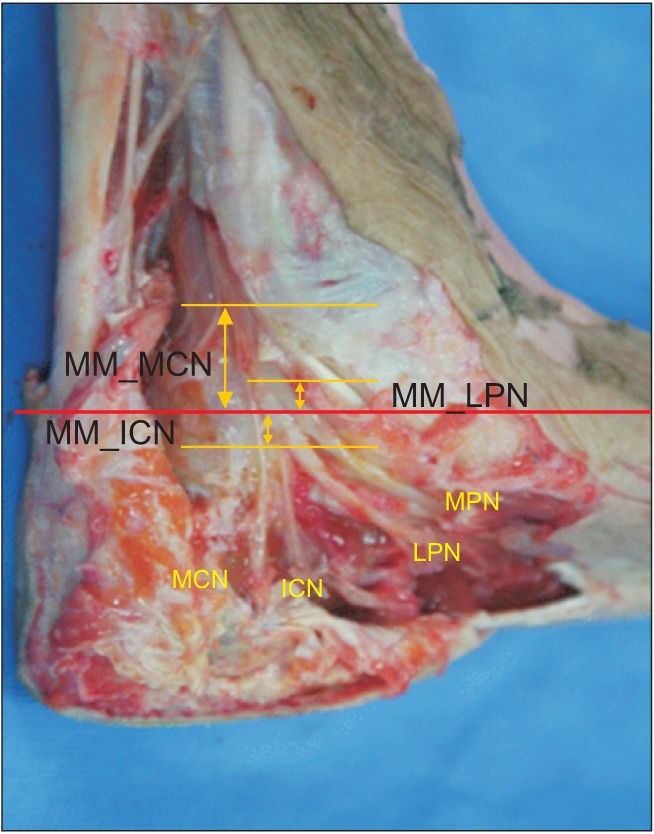
Fig. 1 The distances between the reference line and the bifurcation points were measured. MM, medial malleolus; MCN, medial calcaneal nerve; MPN, medial plantar nerve; LPN, lateral plantar nerve; ICN, inferior calcaneal nerve; MM_MCN, distance between the reference line and the bifurcation point to medial calcaneal nerve; MM_ICN, distance between the reference line and the bifurcation point to inferior calcaneal nerve; MM_LPN, distance between the reference line and the bifurcation point to lateral plantar nerve.
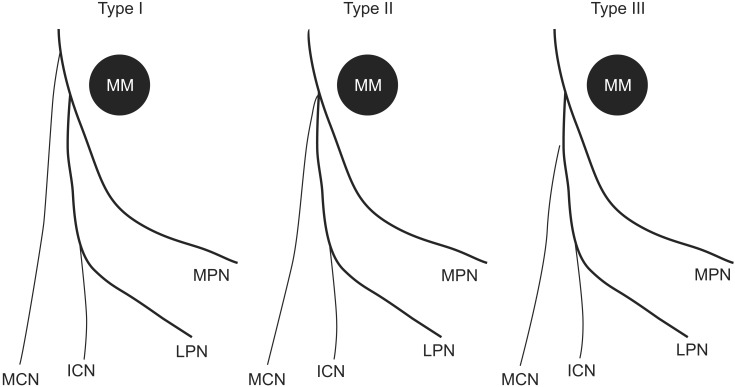
Fig. 2 The branching patterns of tibial nerve were categorized into three types as described in the text. MM, medial malleolus; MCN, medial calcaneal nerve; MPN, medial plantar nerve; LPN, lateral plantar nerve; ICN, inferior calcaneal nerve.
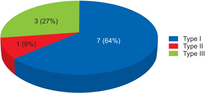
Fig. 3 The bifurcation patterns for tibial nerve. Type I, a pattern in which the medial calcaneal nerve branched proximal to the bifurcation point of medial and lateral plantar nerves; type II, a branching pattern in which the three branches occurred at the same point; type III, a pattern in which the medial calcaneal nerve branched distal to the bifurcation point of the medial and lateral plantar nerves.
Cited by 1 articles
-
Anatomical study and branching point of neurovascular structures at the medial side of the ankle
Chanatporn Inthasan, Tanawat Vaseenon, Pasuk Mahakkanukrauh
Anat Cell Biol. 2020;53(4):422-434. doi: 10.5115/acb.20.087.
Reference
-
1. Williams PL, Warwick R, Dyson M, Bannister LH. Gray's anatomy: the anatomical basis of medicine and surgery. 38th ed. Philadelphia: Saunders;1995.2. Moore KL, Dalley AF. Clinically oriented anatomy. 4th ed. Philadelphia: Lippincott Williams & Wilkins;1999.3. Draves DJ. Anatomy of the lower extremity. 6th ed. Baltimore: Williams & Wilkins;1986.4. Ellis H. Clinical anatomy. 8th ed. Oxford: Blackwell Scientific Publications;1992.5. Gardner ED, Gray DJ, O'Rahilly R. Anatomy: a regional study of human structure. 3rd ed. Philadelphia: Saunders;1969.6. Bareither DJ, Genau JM, Massaro JC. Variation in the division of the tibial nerve: application to nerve blocks. J Foot Surg. 1990; 29:581–583. PMID: 2292650.7. Thapa D, Ahuja V. Combination of diagnostic medial calcaneal nerve block followed by pulsed radiofrequency for plantar fascitis pain: a new modality. Indian J Anaesth. 2014; 58:183–185. PMID: 24963184.
Article8. Martin-Oliva X, Elgueta-Grillo J, Veliz-Ayta P, Orosco-Villasenor S, Elgueta-Grillo M, Viladot-Perice R. Anatomical variants of the medial calcaneal nerve and the Baxter nerve in the tarsal tunnel. Acta Ortop Mex. 2013; 27:38–42. PMID: 24701749.9. Santi MD, Botte MJ. External fixation of the calcaneus and talus: an anatomical study for safe pin insertion. J Orthop Trauma. 1996; 10:487–491. PMID: 8892149.
Article10. Casey D, McConnell T, Parekh S, Tornetta P 3rd. Percutaneous pin placement in the medial calcaneus: is anywhere safe? J Orthop Trauma. 2002; 16:26–29. PMID: 11782629.
Article11. Mekhail AO, Ebraheim NA, Heck BE, Yeasting RA. Anatomic considerations for safe placement of calcaneal pins. Clin Orthop Relat Res. 1996; (332):254–259. PMID: 8913170.
Article12. Greene DL, Thompson MC, Gesink DS, Graves SC. Anatomic study of the medial neurovascular structures in relation to calcaneal osteotomy. Foot Ankle Int. 2001; 22:569–571. PMID: 11503981.
Article13. Horwitz MT. Normal anatomy and variations of the peripheral nerves of the leg and foot. Arch Surg. 1938; 36:626–636.
Article
- Full Text Links
-
- Actions
-
Cited
- CITED
-
- Close
- Share
-
- Similar articles
-
- Distal Motor Nerve Conduction Studies of Medial Plantar Nerve, Lateral Plantar Nerve and Inferior Calcaneal Nerve
- Anatomical study and branching point of neurovascular structures at the medial side of the ankle
- Tarsal Tunnel Syndrome: A Case Report
- Surgical Decompression of Tarsal Tunnel Syndrome Associated with Ganglion Cyst: A Case Report
- Operative Treatment of the Tarsal Tunnel Syndrome Caused by Tarsal Coalition
