Painful Os Peroneum Syndrome Presenting as Lateral Plantar Foot Pain
- Affiliations
-
- 1Department of Rehabilitation Medicine, Incheon St. Mary's Hospital, The Catholic University of Korea, Incheon 403-720, Korea. minukkim@nate.com
- 2Department of Radiology, Incheon St. Mary's Hospital, The Catholic University of Korea, Incheon 403-720, Korea.
- 3Department of Orthopedic Surgery, Seoul St. Mary's Hospital, The Catholic University of Korea, Seoul 137-701, Korea.
- KMID: 2266797
- DOI: http://doi.org/10.5535/arm.2012.36.1.163
Abstract
- Lateral plantar foot pain can be caused by various entities, and the painful os peroneum syndrome should be considered in the differential diagnosis. Recent developments in musculoskeletal ultrasonography are very useful for initial diagnosis. We discuss a 69-year-old female who experienced lateral plantar foot pain for over one month. Through physical examination, radiography, ultrasound and magnetic resonance imaging, she was diagnosed with the painful os peroneum syndrome with a chronic fatigue fracture of multipartite os peroneum and peroneus longus tenosynovitis, for which she underwent surgery. We herein report this rare condition and reviewed the relevant literature.
MeSH Terms
Figure
-
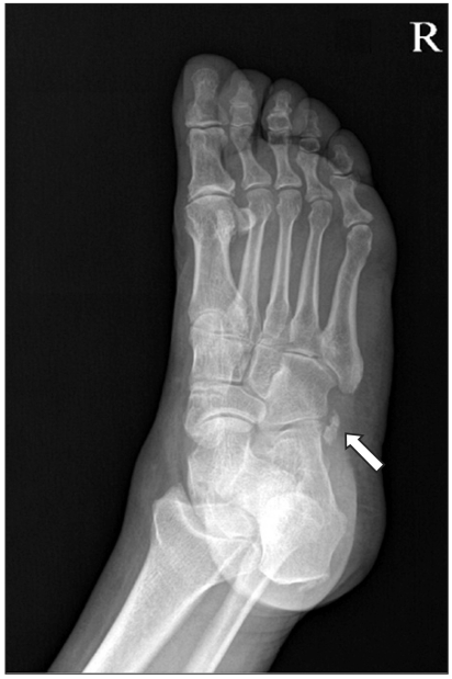
Fig. 1 Oblique view of the foot shows multipartite os peroneum at the level of the calcaneocuboid articulation (arrow).
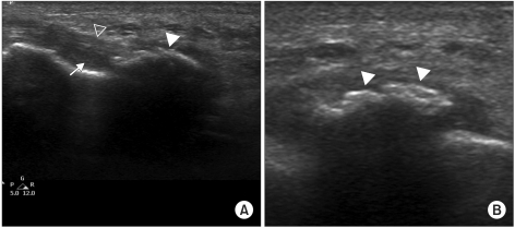
Fig. 2 Longitudinal ultrasonography view shows tenosynovitis (arrow) of the peroneus longus tendon (empty arrowhead) aligned to os peroneum (arrowhead)(A), and transverse ultrasonography view shows cortical disruption of the os peroneum (arrowheads) (B).
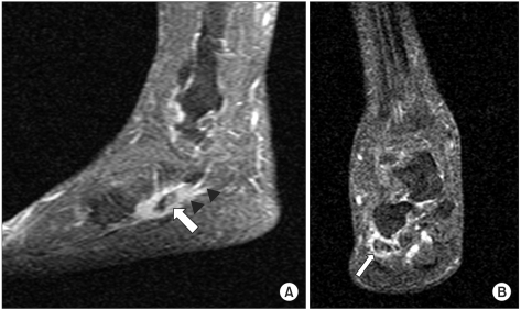
Fig. 3 Sagittal (A) and coronal (B) fat-suppressed contrast-enhanced T1-weighted image (TR/TE 780/12) demonstrates os peroneum (arrow) near the cuboid bone, which is in the inflamed peroneus longus tendon (arrowheads).
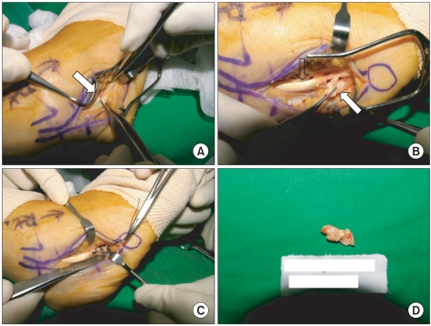
Fig. 4 Intraoperative photograph shows an os peroneum (arrow) in the peroneus longus tendon (A), peroneus longus tendon (empty arrow), and excision of multipartite os peroneum (arrow) (B), anastomosis of peroneus longus tendon (C), and the excised multipartite os peroneum (D).
Reference
-
1. Benjamin M, Qin S, Ralphs JR. Fibrocartilage associated with human tendons and their pulleys. J Anat. 1995; 187:625–633. PMID: 8586561.2. Le Minor JM. Comparative anatomy and significance of the sesamoid bone of the peroneus longus muscle (os peroneum). J Anat. 1987; 151:85–99. PMID: 3654363.3. Coskun N, Yuksel M, Cevener M, Arican RY, Ozdemir H, Bircan O, Sindel T, Ilgi S, Sindel M. Incidnece of accessory ossicles and sesamoid bones in the feet: a radiographic study of the Turkish subjects. Surg Radiol Anat. 2009; 31:19–24. PMID: 18633564.4. Muehleman C, Williams J, Bareither ML. A radiologic and histologic study of the os peroneum: prevalence, morphology, and relationship to degenerative joint disease of the foot and ankle in a cadaveric sample. Clin Anat. 2009; 22:747–754. PMID: 19637293.
Article5. Sobel M, Pavlov H, Geppert MJ, Thompson FM, DiCarlo EF, Davis WH. Painful os peroneum syndrome: a spectrum of conditions responsible for plantar lateral foot pain. Foot Ankle Int. 1994; 15:112–124. PMID: 7951939.
Article6. Bae SY, Chung HJ, Oh JS. Fracture of os peroneum with rupture of the peroneus longus tendon-a case report. J Korean Foot Ankle Soc. 2009; 13:207–210.7. Kim YH, Kim KW, Min HJ, Yoon ES, Kim HO, Suh JS. Fracture of the os peroneumwith rupture of the peroneus longus tendon-a case report. J Korean Soc Fract. 2001; 14:685–688.
- Full Text Links
-
- Actions
-
Cited
- CITED
-
- Close
- Share
-
- Similar articles
-
- Fracture of Os Peroneum with Rupture of the Peroneus Longus Tendon: A Case Report
- Total Rupture of Peroneus Longus Tendon Through an Os Peroneum Fracture Treated by Tendon Transfer (A Case Report)
- Fracture of the Os Peroneum with Rupture of the Peroneus Longus Tendon: A Case Report
- Plantar Fibromatosis: A Case Report
- Treatment of Os Trigonum Syndrome using Subtalar Arthroscopy: A Case Report
