Ann Dermatol.
2010 Nov;22(4):456-459. 10.5021/ad.2010.22.4.456.
Atrophying Pityriasis Versicolor: Is This a New Variant of Pityriasis Versicolor?
- Affiliations
-
- 1Department of Dermatology, School of Medicine, Kyung Hee University, Seoul, Korea. crhaw@khmc.or.kr
- KMID: 2266187
- DOI: http://doi.org/10.5021/ad.2010.22.4.456
Abstract
- An atypical clinical form of pityriasis versicolor has been infrequently reported, in which cutaneous atrophy is associated with individual pityriasis versicolor lesions. The pathogenesis of this atrophy remains unclear, but is believed to be a delayed-type hypersensitivity reaction to antigens derived from the Malassezia species. A 60-year-old man presented with multiple, slightly scaly, and depressed maculopatches or plaques on the trunk and extremities. Our microscopic examination of the skin scrapings on a KOH preparation revealed numerous short hyphae and spores. The patient was treated daily with 200 mg of itraconazole in combination with topical antifungals, achieving clinical improvement and mycological recovery, which was confirmed upon follow-up 1 month later. This is the first case report of atrophying pityriasis versicolor in Korea. It needs to be differentiated from other atrophying disorders of the skin.
Keyword
MeSH Terms
Figure
-
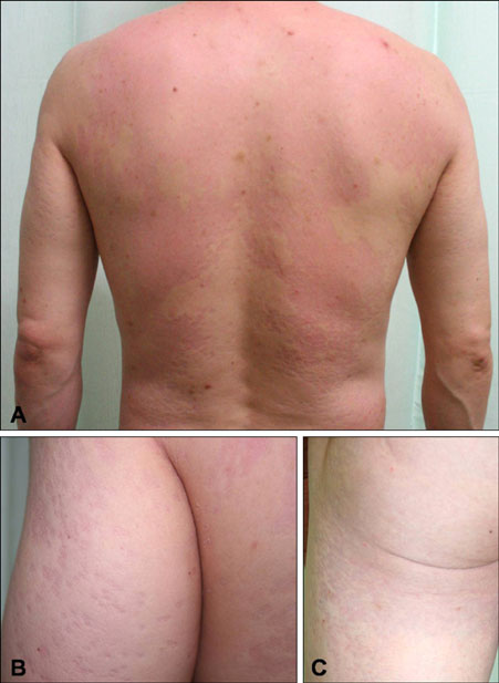
Fig. 1 Numerous coin-sized confluent erythematous atrophic patches or plaques with scales on the back (A), upper arm (B), and buttock and thigh (C).
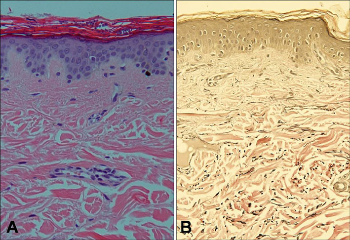
Fig. 2 (A) Multiple roundish spores with some hyphae of Malassezia in the horny layer and focal thinning of the epidermis (H&E, ×200). (B) Focal decreased and fragmented elastic fibers (Verhoff-van Gieson, ×200).
Reference
-
1. Gupta AK, Batra R, Bluhm R, Faergemann J. Pityriasis versicolor. Dermatol Clin. 2003. 21:413–429. v–vi.
Article2. Crowson AN, Magro CM. Atrophying tinea versicolor: a clinical and histological study of 12 patients. Int J Dermatol. 2003. 42:928–932.
Article3. De Graciansky P, Mery F. Atrophie sur pityriasis versicolor apres corticotherapie locale prolongee. Bull Soc Fr Dermatol Syphiligr. 1971. 78:295.4. Schöpf E. Adverse effects of external corticosteroid therapy. Hautarzt. 1972. 23:295–301.5. Runne U. Pseudo-Anetodermie im Bereich einer mit Corticosteroidexterna behandelten Pityriasis versicolor (2 Falle). Z Hautkr. 1975. 50:449–451.6. Mascaro JM, Torres V. Pitiriasis versicolor atrofica. Dermatologia. 1976. 20:329–336.7. Paradisi M, Constantini G, Muscianese V, Voglino A. Pityriasis versicolor atrophicans. Chron Derm. 1978. 5:564–565.8. Millaseca ML, Gonzalez-Herrada C, Herranz JM, Corripio F, Jaquetti G. Pitiriasis versicolor atrofica yatrogenica. Gaceta Dermatologica. 1981. 2:108–112.9. Tatnall FM, Rycroft RJ. Pityriasis versicolor with cutaneous atrophy induced by topical steroid application. Clin Exp Dermatol. 1985. 10:258–261.
Article10. Wagner G, Lubach D. Pityriasis versicolor pseudoatrophicans. A case description. Z Hautkr. 1987. 62:321–324.11. Vera Castano A, Trasobares Marugan L, Del valle Martin M, Hernanz Hermosa JM, Del Palacio Hernanz A. Pitiriasis versicolor atrofica inducida por corticoides topicos fluorados. Actas Dermo-Sif. 1989. 80:69–71.12. Mazuecos Blanca J, García-Bravo B, Moreno Giménez JC, Sotillo I, Camacho F. Pseudoatrophic pityriasis versicolor. Med Cutan Ibero Lat Am. 1990. 18:101–103.13. Fimiani M, Casini L, Simoni S, Andreassi A, Alessandrini C. Un caso di pitiriasi versicolor atrofica: aspetti ultrastrutturali. Micol Dermatol. 1992. 6:21–30.14. Romano C, Maritati E, Ghilardi A, Miracco C, Mancianti F. A case of pityriasis versicolor atrophicans. Mycoses. 2005. 48:439–441.
Article15. Winter GD, Burton JL. Experimentally induced steroid atrophy in the domestic pig and man. Br J Dermatol. 1976. 94:suppl 12. 107–109.
Article16. Tosti A, Villardita S, Fazzini ML. The parasitic colonization of the horny layer in tinea versicolor. J Invest Dermatol. 1972. 59:233–237.
Article17. Lehmann P, Zheng P, Lavker RM, Kligman AM. Corticosteroid atrophy in human skin. A study by light, scanning, and transmission electron microscopy. J Invest Dermatol. 1983. 81:169–176.
Article18. Jablonska S, Groniowska M, Dabroswki J. Comparative evaluation of skin atrophy in man induced by topical corticoids. Br J Dermatol. 1979. 100:193–206.
Article
- Full Text Links
-
- Actions
-
Cited
- CITED
-
- Close
- Share
-
- Similar articles
-
- Clinical Efficacy and Tolerability of Terbinafine 1% Cream in Patients with Pityriasis Versicolor
- Atrophying Pityriasis Versicolor
- Pityriasis Versicolor Atrophicans
- Two Cases of Pityriasis Versicolor on the Scalp in the Course of Treatment for Alopecia Totalis
- Malassezia Species Cultured from the Lesions of Pityriasis Versicolor
