Ann Dermatol.
2012 Nov;24(4):480-481. 10.5021/ad.2012.24.4.480.
Adult Onset of Nevus Unius Lateris
- Affiliations
-
- 1Department of Dermatology, Chung-Ang University College of Medicine, Seoul, Korea. drseo@hanafos.com
- KMID: 2266050
- DOI: http://doi.org/10.5021/ad.2012.24.4.480
Abstract
- No abstract available.
Figure
-
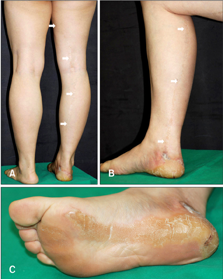
Fig. 1 (A, B, C) The skin lesions appeared in a linear fashion, following the lines of Blaschko, involving primarily the right leg, which were extended to ipsilateral sole. Right sole manifest as yellowish, thick, linear distributed, verrucous, and hyperkeratotic plaque.

Fig. 2 (A) A biopsy specimen from the right calf shows hyperkeratosis, acanthosis and hyperpigmentation of the basal layer (H&E stain, ×100). (B) Histology performed on a biopsy specimen from a keratotic lesion on the sole reveals verruciform epidermal changes, such as marked hyperkeratosis, parakeratosis and acanthosis (H&E stain, ×100).
Reference
-
1. Happle R, Rogers M. Epidermal nevi. Adv Dermatol. 2002. 18:175–201.2. Haberland-Carrodeguas C, Allen CM, Lovas JG, Hicks J, Flaitz CM, Carlos R, et al. Review of linear epidermal nevus with oral mucosal involvement--series of five new cases. Oral Dis. 2008. 14:131–137.3. Su WP. Histopathologic varieties of epidermal nevus. A study of 160 cases. Am J Dermatopathol. 1982. 4:161–170.
Article4. Mazereeuw-Hautier J, Thibaut I, Bonafé JL. Acantholytic dyskeratotic epidermal nevus: a rare histopathologic feature. J Cutan Pathol. 2002. 29:52–54.
Article5. Fox BJ, Lapins NA. Comparison of treatment modalities for epidermal nevus: a case report and review. J Dermatol Surg Oncol. 1983. 9:879–885.
Article
