Ann Dermatol.
2014 Feb;26(1):121-122. 10.5021/ad.2014.26.1.121.
Congenital Form of Isolated Benign Primary Cutaneous Plasmacytosis in a Child
- Affiliations
-
- 1Department of Dermatology, Ajou University School of Medicine, Suwon, Korea. maychan@ajou.ac.kr
- KMID: 2265713
- DOI: http://doi.org/10.5021/ad.2014.26.1.121
Abstract
- No abstract available.
Figure
-
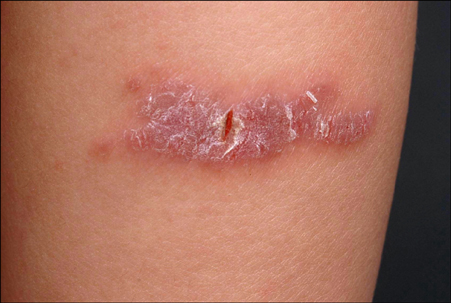
Fig. 1 A solitary asymptomatic erythematous plaque on the right lower leg.

Fig. 2 (A) Psoriasiform hyperplasia with dense lymphoplasma cells infiltration in the superficial and deep dermis without atypia (H&E, ×10). Polyclonal hypergammaglobulinemia was proven with Kappa light chain stain (B) and lambda light chain (C) (immunoperoxidase stain, ×200).
Reference
-
1. Carey WP, Rico MJ, Nierodzik M, Sidhu G. Systemic plasmacytosis with cutaneous manifestations in a white man: successful therapy with cyclophosphamide/prednisone. J Am Acad Dermatol. 1998; 38:629–631.
Article2. Aricò M, Bongiorno MR. Primary cutaneous plasmacytosis in a child. Is this a new entity? J Eur Acad Dermatol Venereol. 2002; 16:164–167.
Article3. Jayaraman AG, Cesca C, Kohler S. Cutaneous plasmacytosis: A report of five cases with immunohistochemical evaluation for HHV-8 expression. Am J Dermatopathol. 2006; 28:93–98.4. Leonard AL, Meehan SA, Ramsey D, Brown L, Sen F. Cutaneous and systemic plasmacytosis. J Am Acad Dermatol. 2007; 56:2 Suppl. S38–S40.
Article5. Ahn JJ, Yang YS, Shin MK, Lee SW, Kim NI. Case of isolated benign primary cutaneous plasmacytosis in a child. J Dermatol. 2011; 38:364–367.
Article
