Ann Dermatol.
2014 Feb;26(1):115-116. 10.5021/ad.2014.26.1.115.
Nasal Sinus Tract Associated with Dental Infection
- Affiliations
-
- 1Department of Clinical and Preventive Dentistry, Federal University of Pernambuco, Pernambuco, Brazil. danyel.perez@ufpe.br
- 2Department of Stomatology, A. C. Camargo Hospital, Sao Paulo, Brazil.
- KMID: 2265710
- DOI: http://doi.org/10.5021/ad.2014.26.1.115
Abstract
- No abstract available.
Figure
-

Fig. 1 (A) An erythematous painless nodule in the right nostril. (B) A diffuse radiolucent image in the periapical region of the right maxillary central incisor, with disruption of the lamina dura (cortical alveolar bone) (arrows).
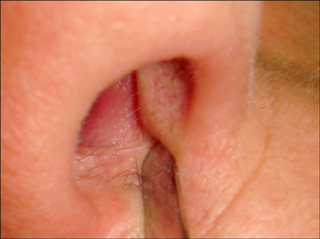
Fig. 2 Complete healing of the nasal lesion after the nonsurgical endodontic treatment.
Reference
-
1. Pasternak-Júnior B, Teixeira CS, Silva-Sousa YT, Sousa-Neto MD. Diagnosis and treatment of odontogenic cutaneous sinus tracts of endodontic origin: three case studies. Int Endod J. 2009; 42:271–276.
Article2. Fowler EB, Breault LG, Galvan DA. Nasal fistula associated with dental infection: a report of a case. J Endod. 2000; 26:374–376.
Article3. Güleç AT, Seçkin D, Bulut S, Sarfakoğlu E. Cutaneous sinus tract of dental origin. Int J Dermatol. 2001; 40:650–652.
Article4. Cohen PR, Eliezri YD. Cutaneous odontogenic sinus simulating a basal cell carcinoma: case report and literature review. Plast Reconstr Surg. 1990; 86:123–127.5. Michaelson PL, Holland GR. Is pulpitis painful? Int Endod J. 2002; 35:829–832.
Article
