Ann Dermatol.
2014 Aug;26(4):545-546. 10.5021/ad.2014.26.4.545.
Lipomatosis of the Nerves in the Back
- Affiliations
-
- 1Department of Dermatology, CHA Bundang Medical Center, CHA University, Seongnam, Korea. msch11@chamc.co.kr
- KMID: 2265609
- DOI: http://doi.org/10.5021/ad.2014.26.4.545
Abstract
- No abstract available.
MeSH Terms
Figure
-
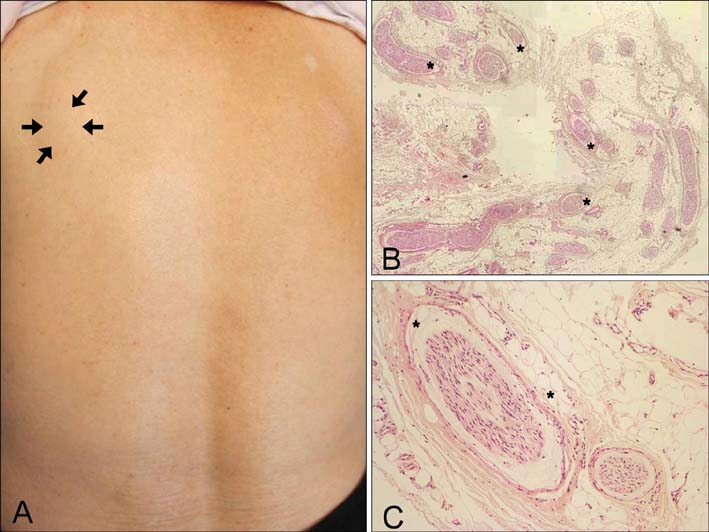
Fig. 1 (A) A flesh-colored subcutaneous mass (arrows) was noticed on the left posterior scapula area. (B) A punch biopsy specimen revealed an increased amount of adipose tissue (asterisks) around nerve bundles in the deep subcutaneous fat layer (H&E, ×40). (C) Enlargement of the nerve bundles due to fibrous thickening and an increased amount of adipose tissue (asterisks) (H&E, ×200).
Reference
-
1. Bancroft LW, Kransdorf MJ, Peterson JJ, O'Connor MI. Benign fatty tumors: classification, clinical course, imaging appearance, and treatment. Skeletal Radiol. 2006; 35:719–733.
Article2. Murphey MD, Smith WS, Smith SE, Kransdorf MJ, Temple HT. From the archives of the AFIP. Imaging of musculoskeletal neurogenic tumors: radiologic-pathologic correlation. Radiographics. 1999; 19:1253–1280.3. Venkatesh K, Saini ML, Rangaswamy R, Murthy S. Neural fibrolipoma without macrodactyly: a subcutaneous rare benign tumor. J Cutan Pathol. 2009; 36:594–596.
Article
