Ann Dermatol.
2014 Aug;26(4):501-504. 10.5021/ad.2014.26.4.501.
Two Concurrent Facial Epidermal Nevi without Systemic Abnormalities: Nevus Sebaceus and Nevus Comedonicus
- Affiliations
-
- 1Department of Dermatology, Kyungpook National University School of Medicine, Daegu, Korea. weonju@knu.ac.kr
- KMID: 2265593
- DOI: http://doi.org/10.5021/ad.2014.26.4.501
Abstract
- Epidermal nevi (EN) are hamartomatous lesions derived from epidermal components originating from pluripotent cell mutations. They have been categorized according to their predominant component. The existence of >2 types of EN concurrently within a single area or within contiguous areas has been rarely reported. This report describes the case of simultaneous presence of a yellowish plaque on the left medial canthus and an aggregation of closed comedo-like papules on the right side of the cheek of a 15-year-old girl.
Keyword
MeSH Terms
Figure
-
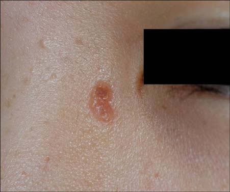
Fig. 1 A yellowish, bean-sized, verrucous plaque on the left medial canthus, and multiple, scattered, pea-sized, hyperpigmented macules on the face.
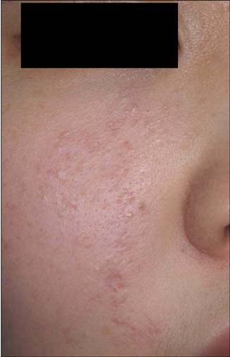
Fig. 2 An aggregation of closed comedo-like papules and depressed pinpoint scars on the right side of the cheek.
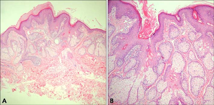
Fig. 3 (A) Marked acanthotic and papillomatous epidermal hyperplasia with hyperkeratosis (H&E, ×40). (B) Excessive number of large sebaceous glands (H&E, ×100).
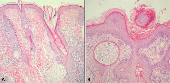
Fig. 4 (A) A wide, deep, epidermal invagination filled with keratin and incomplete hair follicles (H&E, ×100). (B) Well-defined keratin-filled cyst in the mid-dermis (H&E, ×100).
Reference
-
1. Vidaurri-de la Cruz H, Tamayo-Sánchez L, Durán-McKinster C, de la Luz Orozco-Covarrubias M, Ruiz-Maldonado R. Epidermal nevus syndromes: clinical findings in 35 patients. Pediatr Dermatol. 2004; 21:432–439.
Article2. Solomon LM, Esterly NB. Epidermal and other congenital organoid nevi. Curr Probl Pediatr. 1975; 6:1–56.
Article3. Rogers M, McCrossin I, Commens C. Epidermal nevi and the epidermal nevus syndrome. A review of 131 cases. J Am Acad Dermatol. 1989; 20:476–488.4. Hodge JA, Ray MC, Flynn KJ. The epidermal nevus syndrome. Int J Dermatol. 1991; 30:91–98.
Article5. Moss C, Larkins S, Stacey M, Blight A, Farndon PA, Davison EV. Epidermal mosaicism and Blaschko's lines. J Med Genet. 1993; 30:752–755.
Article6. Rogers M. Epidermal nevi and the epidermal nevus syndromes: a review of 233 cases. Pediatr Dermatol. 1992; 9:342–344.
Article7. Muñoz-Pérez MA, García-Hernandez MJ, Ríos JJ, Camacho F. Sebaceous naevi: a clinicopathological study. J Eur Acad Dermatol Venereol. 2002; 16:319–324.8. Kofmann S. Ein Fall von seltener Lokalisation und Verbreitung von Komedon. Arch Dermatol Syphilol. 1895; 32:177–178.9. Beck MH, Dave VK. Extensive nevus comedonicus. Arch Dermatol. 1980; 116:1048–1050.
Article10. Decherd JW, Mills O, Leyden JJ. Naevus comedonicus--treatment with retinoic acid. Br J Dermatol. 1972; 86:528–529.11. Köse O, Calişkan E, Kurumlu Z. Three different epidermal naevi with no organ involvement: sebaceous naevus, naevus comedonicus and Becker's naevus. Acta Derm Venereol. 2008; 88:67–69.
Article12. Kim SC, Kang WH. Nevus comedonicus associated with epidermal nevus. J Am Acad Dermatol. 1989; 21:1085–1088.
Article13. Domonkos AN, Arnold HL, Odom RB. Andrew's disease of the skin. 7th ed. Philadelphia: W.B. Saunders Co.;1982. p. 795.14. Hill VA, Felix RH, Mortimer PS, Harper JI. Phacomatosis pigmentokeratotica. J R Soc Med. 2003; 96:30–31.
Article15. Happle R, Hoffmann R, Restano L, Caputo R, Tadini G. Phacomatosis pigmentokeratotica: a melanocytic-epidermal twin nevus syndrome. Am J Med Genet. 1996; 65:363–365.
Article
- Full Text Links
-
- Actions
-
Cited
- CITED
-
- Close
- Share
-
- Similar articles
-
- A Case of Verrucous Epidermal Nevus Contiguous with Nevus Sebaceus
- Nevus Comedonicus Associated with Epidermal Cyst
- A Case of nevus Sebacens of Jadassohn Associated with Various Epidermal Appendage Tumors
- Keratoacanthoma in Co-existence with Nevus Sebaceus
- A Case of Nevus Sebaceus Syndrome Considered as a Neurocutaneous Syndrome
