Keloids and Hypertrophic Scars: Characteristic Vascular Structures Visualized by Using Dermoscopy
- Affiliations
-
- 1Department of Dermatology, Korea University College of Medicine, Seoul, Korea. kumcihk@korea.ac.kr
- KMID: 2265494
- DOI: http://doi.org/10.5021/ad.2014.26.5.603
Abstract
- BACKGROUND
Keloids and hypertrophic scars represent excessive scarring. They require different therapeutic approaches, which can be hampered because of an apparent lack of morphologic difference between the two diseases.
OBJECTIVE
This study investigated the clinical and dermoscopic features of keloids and hypertrophic scars in order to help dermatologists distinguish these lesions better.
METHODS
A total of 41 keloids and hypertrophic scars in 41 patients were examined clinically and by performing dermoscopy with a digital imaging system. Lesions were evaluated for vascular structures.
RESULTS
Dermoscopy revealed vascular structures in most keloid lesions (90%) but in only 27% of hypertrophic scar lesions. The most common dermoscopic vascular structures in keloids were arborizing (52%), followed by linear irregular (33%) and commashaped (15%); these features were present but less evident in hypertrophic scars (9% for all types). The distribution frequency of the vascular structures differed significantly between diseases (p<0.001).
CONCLUSION
A strong association of vascular structures with keloids was observed on dermoscopic examination. The results suggest dermoscopic examination of vascular structures is a clinically useful diagnostic tool for differentiating between keloids and hypertrophic scars.
Figure
-
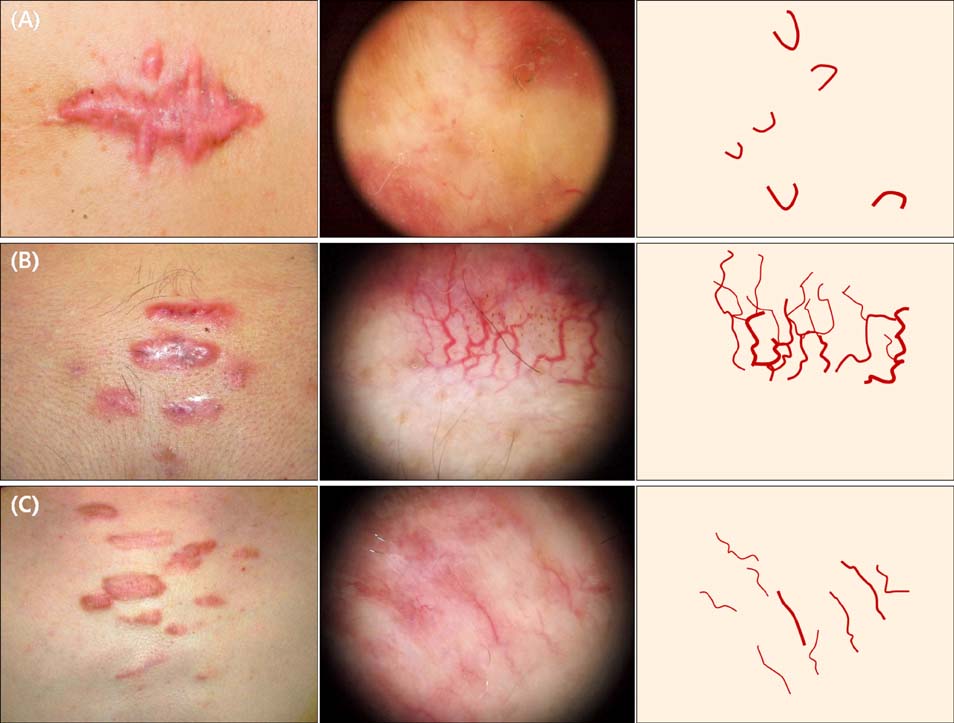
Fig. 1 Clinical and dermoscopic findings with schematic drawing of the morphological types of vascular structures of patients with keloids. (A) Large erythematous hard nodules on the chest; comma-shaped vessels are visible. (B) Multiple erythematous hard nodules on the chest; arborizing vessels are visible. (C) Multiple erythematous hard nodules on the chest; linear irregular vessels are visible.
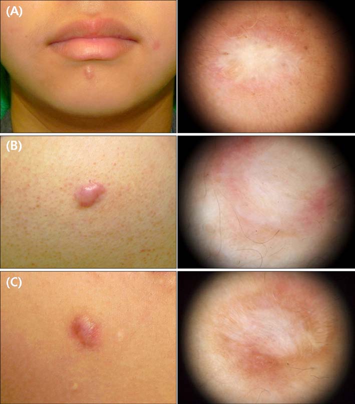
Fig. 2 Clinical and dermoscopic findings of patients with hypertrophic scars. (A) Dome-shaped hard nodules on the chin; no vascular structures are apparent. (B) Dome-shaped erythematous hard nodules on the back; no vascular structures are apparent. (C) Dome-shaped erythematous hard nodules on the chest; no vascular structures are apparent.
Cited by 1 articles
-
Arborizing Vessels on Dermoscopy in Various Skin Diseases Other Than Basal Cell Carcinoma
Hyunju Jin, Min-Young Yang, Jeong-Min Kim, Gun-Wook Kim, Hoon-Soo Kim, Hyun-Chang Ko, Byung-Soo Kim, Moon-Bum Kim
Ann Dermatol. 2017;29(3):288-294. doi: 10.5021/ad.2017.29.3.288.
Reference
-
1. English RS, Shenefelt PD. Keloids and hypertrophic scars. Dermatol Surg. 1999; 25:631–638.
Article2. Craig RD, Schofield JD, Jackson DS. Collagen biosynthesis in normal and hypertrophic scars and keloid as a function of the duration of the scar. Br J Surg. 1975; 62:741–744.
Article3. Köse O, Waseem A. Keloids and hypertrophic scars: are they two different sides of the same coin? Dermatol Surg. 2008; 34:336–346.
Article4. Bran GM, Goessler UR, Hormann K, Riedel F, Sadick H. Keloids: current concepts of pathogenesis (review). Int J Mol Med. 2009; 24:283–293.
Article5. Campos-do-Carmo G, Ramos-e-Silva M. Dermoscopy: basic concepts. Int J Dermatol. 2008; 47:712–719.
Article6. Roques C, Téot L. The use of corticosteroids to treat keloids: a review. Int J Low Extrem Wounds. 2008; 7:137–145.
Article7. Gauglitz GG, Korting HC, Pavicic T, Ruzicka T, Jeschke MG. Hypertrophic scarring and keloids: pathomechanisms and current and emerging treatment strategies. Mol Med. 2011; 17:113–125.
Article8. Zalaudek I, Kreusch J, Giacomel J, Ferrara G, Catricalà C, Argenziano G. How to diagnose nonpigmented skin tumors: a review of vascular structures seen with dermoscopy: part I. Melanocytic skin tumors. J Am Acad Dermatol. 2010; 63:361–374.
Article9. Ruocco E, Argenziano G, Pellacani G, Seidenari S. Noninvasive imaging of skin tumors. Dermatol Surg. 2004; 30:301–310.
Article10. Bouzari N, Davis SC, Nouri K. Laser treatment of keloids and hypertrophic scars. Int J Dermatol. 2007; 46:80–88.
Article11. Ehrlich HP, Desmoulière A, Diegelmann RF, Cohen IK, Compton CC, Garner WL, et al. Morphological and immunochemical differences between keloid and hypertrophic scar. Am J Pathol. 1994; 145:105–113.12. Datubo-Brown DD. Keloids: a review of the literature. Br J Plast Surg. 1990; 43:70–77.
Article13. Babu M, Bai RP, Suguna L, Ramachandran K, Ramakrishnan KM. Differentiation of keloid and hypertrophic scar; correlation of the water proton relaxation times with the duration of the scar. Physiol Chem Phys Med NMR. 1993; 25:113–120.14. Kischer CW, Shetlar MR, Chvapil M. Hypertrophic scars and keloids: a review and new concept concerning their origin. Scan Electron Microsc. 1982; 1699–1713.15. Le AD, Zhang Q, Wu Y, Messadi DV, Akhondzadeh A, Nguyen AL, et al. Elevated vascular endothelial growth factor in keloids: relevance to tissue fibrosis. Cells Tissues Organs. 2004; 176:87–94.
Article16. Lametschwandtner A, Staindl O. Angioarchitecture of keloids. A scanning electron microscopy study of a corrosion specimen. HNO. 1990; 38:202–207.17. Lee JY, Yang CC, Chao SC, Wong TW. Histopathological differential diagnosis of keloid and hypertrophic scar. Am J Dermatopathol. 2004; 26:379–384.
Article18. Atiyeh BS, Costagliola M, Hayek SN. Keloid or hypertrophic scar: the controversy: review of the literature. Ann Plast Surg. 2005; 54:676–680.19. Bayat A, Bock O, Mrowietz U, Ollier WE, Ferguson MW. Genetic susceptibility to keloid disease: transforming growth factor beta receptor gene polymorphisms are not associated with keloid disease. Exp Dermatol. 2004; 13:120–124.
Article
- Full Text Links
-
- Actions
-
Cited
- CITED
-
- Close
- Share
-
- Similar articles
-
- A Study on the Effects of 595nm Pulsed Dye Laser Treatment on Scars and Keloids
- Bleomycin Intralesional Injection in Keloids and Hypertrophic Scars Unresponsive to Previous Corticosteroid Intralesional Injection and/or Laser Treatment: A Case Series and Review of the Literature
- Pilot Study of the Efficacy of 578 nm Copper Bromide Laser Combined with Intralesional Corticosteroid Injection for Treatment of Keloids and Hypertrophic Scars
- Effect of Cryosurgery on Hypertrophic Scars / Keloids
- Evaluation of the Skin Barrier Function by TEWL Measurement in Hypertrophic scars and Keloids
