Ann Dermatol.
2010 Aug;22(3):358-361. 10.5021/ad.2010.22.3.358.
Angiolymphoid Hyperplasia with Eosinophilia on the Palm
- Affiliations
-
- 1Department of Dermatology, School of Medicine, Gyeongsang National University, Jinju, Korea.
- 2Institute of Health Sciences, School of Medicine, Gyeongsang National University, Jinju, Korea. jaymax1@dreamwiz.com
- KMID: 2265377
- DOI: http://doi.org/10.5021/ad.2010.22.3.358
Abstract
- Angiolymphoid hyperplasia with eosinophilia (ALHE) is an uncommon dermal angioproliferating tumor, characterized by red to brown papules or nodules on the head and neck, though also occurring in the mouth, trunk, extremities and inguinal area. The palm is a very unusual site for ALHE, and there have been very few cases reported globally thus far. ALHE can be pruritic and painful and histopathologic findings show vascular proliferation with infiltration of eosinophils and lymphocytes in the dermis. Plump endothelial cells protrude into the lumen. We report a case of ALHE occurring at an unusual site, the right palm, in a 62-year-old man, who had suffered from a solitary pinkish-colored, central depressed round hyperkeratotic plaque on his palm for 4 years. On the basis of clinical and histopathologic data, a diagnosis of ALHE was made. To our knowledge, this is the first report of ALHE on the palm in Korean dermatologic literature.
MeSH Terms
Figure
-
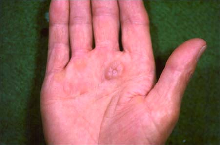
Fig. 1 Solitary, pinkish-colored, central depressed, round hyperkeratotic plaque on the right palm.

Fig. 2 Histopathologic examination demonstrates (A) multiple lobular infiltrations of inflammatory cells with central proliferation of vessels in the dermis (H&E, ×40). (B) Proliferation of blood vessels with infiltration of lymphocytes and eosinophils in the dermis (H&E, ×200). (C) Endothelial cells protruding into the lumen with hobnail appearance (arrowheads) and some vacuolated endothelial cells (arrow) (H&E, ×400).

Fig. 3 Endothelial cells are positive in (A) CD31 (×100) and (B) CD34 (×100).
Reference
-
1. Wells GC, Whimster IW. Subcutaneous angiolymphoid hyperplasia with eosinophilia. Br J Dermatol. 1969. 81:1–14.
Article2. Urabe A, Tsuneyoshi M, Enjoji M. Epithelioid hemangioma versus Kimura's disease. A comparative clinicopathologic study. Am J Surg Pathol. 1987. 11:758–766.3. Kuo TT, Shih LY, Chan HL. Kimura's disease. Involvement of regional lymph nodes and distinction from angiolymphoid hyperplasia with eosinophilia. Am J Surg Pathol. 1988. 12:843–854.4. Lee JS, Lee JB, Kim SJ, Lee SC, Won YH, Yun SJ. A case of angiolymphoid hyperplasia with eosinophilia of the lower eyelid. Ann Dermatol. 2008. 20:138–141.
Article5. Weiss SW, Enzinger FM. Epithelioid hemangioendothelioma: a vascular tumor often mistaken for a carcinoma. Cancer. 1982. 50:970–981.
Article6. Tsang WY, Chan JK. The family of epithelioid vascular tumors. Histol Histopathol. 1993. 8:187–212.7. Olsen TG, Helwig EB. Angiolymphoid hyperplasia with eosinophilia. A clinicopathologic study of 116 patients. J Am Acad Dermatol. 1985. 12:781–796.8. Imbing FD Jr, Viegas SF, Sánchez RL. Multiple angiolymphoid hyperplasia with eosinophilia of the hand: report of a case and review of the literature. Cutis. 1996. 58:345–348.9. Jang KA, Lee JY, Kim CH, Choi JH, Sung KJ, Moon KC, et al. Angiolymphoid hyperplasia with eosinophilia and Kimura's disease: a clinico-pathologic study in Korea. Korean J Dermatol. 2001. 39:309–317.10. Satpathy A, Moss C, Raafat F, Slator R. Spontaneous regression of a rare tumour in a child: angiolymphoid hyperplasia with eosinophilia of the hand: case report and review of the literature. Br J Plast Surg. 2005. 58:865–868.
Article11. Hendricks MW, Moore MM, Dell PC. Angiolymphoid hyperplasia with eosinophilia: a case report. J Hand Surg Am. 1985. 10:286–288.
Article
- Full Text Links
-
- Actions
-
Cited
- CITED
-
- Close
- Share
-
- Similar articles
-
- A Case of Angiolymphoid Hyperplasia with Eosinophilia
- Angiolymphoid Hyperplasia with Eosinophilia in Bone: A Case Report
- A Case of Angiolymphoid Hyperplasia with Eosinophilia
- A Case of Angiolymphoid Hyperplasia with Eosinophilia
- Histiocytoid Hemangioma (Angiolymphoid Hyperplasia with Eosinophilia): Effective Treatment with Dapsone
