A Case of Clear Cell Eccrine Porocarcinoma
- Affiliations
-
- 1Department of Dermatology, School of Medicine, Ewha Womans University, Seoul, Korea. hychoi@ewha.ac.kr
- KMID: 2265370
- DOI: http://doi.org/10.5021/ad.2010.22.3.330
Abstract
- Eccrine porocarcinoma (EP) is a rare malignant tumor arising from the intraepidermal eccrine duct. The tumor cells frequently contain glycogen, but prominent clear cell changes in EP are rarely reported. A 78-year-old woman presented with a slightly pruritic, erythematous, verrucous plaque on her left thigh. Histopathological examination revealed intraepidermal tumor cell nests composed of small basaloid cells and duct-like structures lined by periodic acid-Schiff (PAS)-positive cuticles. Besides the typical findings of EP, clear cell changes were predominantly observed in the tumor cell aggregations. Herein we report a case of the clear cell variant of EP rarely reported in previous literature.
Figure
-
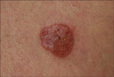
Fig. 1 A 2×2 cm sized, erythematous plaque with focal erosions on the left thigh.

Fig. 2 (A) The intraepidermal aggregations were composed of small basaloid cells with sharp demarcations. Prominent clear cell changes were also noted within the tumor nests (H&E, ×40). (B) Acrosyringium-like, spiral ductal structures were seen in some aggregations (H&E, ×100).
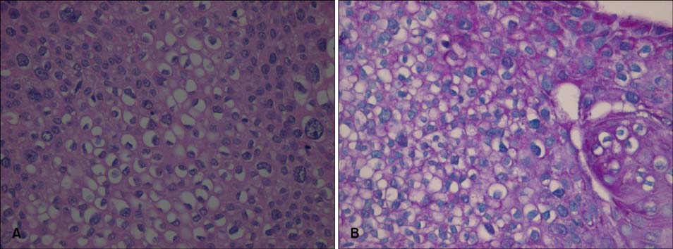
Fig. 3 (A) In addition to atypical tumor cells, numerous clear cells with ample cytoplasm were observed (H&E, ×400). (B) PAS-positive diastase-labile granules were seen in the cytoplasm of clear cells (×400).

Fig. 4 (A) Immunoreactivity for EMA showed focal positivity within the tumor cells (×40). (B) Immunoreactivity for CEA was restricted to the border of the ductal structures (×100).
Reference
-
1. Rutten A, Requena L, Requena C. Clear-cell porocarcinoma in situ: a cytologic variant of porocarcinoma in situ. Am J Dermatopathol. 2002. 24:67–71.2. Pinkus H, Mehregan AH. Epidermotropic eccrine carcinoma. A case combining features of eccrine poroma and Paget's dermatosis. Arch Dermatol. 1963. 88:597–606.3. Robson A, Greene J, Ansari N, Kim B, Seed PT, McKee PH, et al. Eccrine porocarcinoma (malignant eccrine poroma): a clinicopathologic study of 69 cases. Am J Surg Pathol. 2001. 25:710–720.4. Requena L, Sarasa JL, Pique E, Farina MC, Olivares M, Martin L. Clear-cell porocarcinoma: another cutaneous marker of diabetes mellitus. Am J Dermatopathol. 1997. 19:540–544.5. Saitoh A, Ohtake N, Fukuda S, Tamaki K. Clear cells of eccrine glands in a patient with clear cell syringoma associated with diabetes mellitus. Am J Dermatopathol. 1993. 15:166–168.
Article6. Wong TY, Suster S, Nogita T, Duncan LM, Dickersin RG, Mihm MC Jr. Clear cell eccrine carcinomas of the skin. A clinicopathologic study of nine patients. Cancer. 1994. 73:1631–1643.
Article
