A Case of Hydroa Vacciniforme
- Affiliations
-
- 1Department of Dermatology, Samsung Medical Center, Sungkyunkwan University School of Medicine, Seoul, Korea. junmo.yang@samsung.com
- KMID: 2265365
- DOI: http://doi.org/10.5021/ad.2010.22.3.312
Abstract
- Hydroa vacciniforme (HV) is a rare and chronic pediatric disorder that is characterized by photosensitivity and recurrent vesicles that heal with vacciniforme scarring. The pathogenesis of HV is unknown; no chromosome abnormality has been identified. HV patients have no abnormal laboratory results, so the diagnosis of HV is based on identifying the associated histological findings in a biopsy specimen and using repetitive ultraviolet phototesting to reproduce the characteristic vesicles on a patient's skin. Herein, we present a case of HV in a 7-year-old female who was diagnosed with HV according to histopathology and ultraviolet phototesting.
Keyword
Figure
-
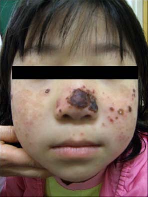
Fig. 1 Erythematous, pitted and atrophic scars with crusting on the nose and cheek.

Fig. 2 Diffuse, hypopigmented and umbilicated scars on the dorsa of the hands.
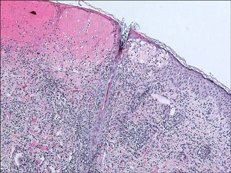
Fig. 3 Histopathology from the erythematous vesicles on the patient's face showed epidermal necrosis, intraepidermal vesicles with spongiosis and a superficial perivascular lymphocytic infiltration (H&E stain, ×100).

Fig. 4 (A) There were no infiltrating mononuclear cells that expressed Epstein-Barr virus-determined nuclear antigens (EBNA) in the lesional skin biopsy specimen (×100). (B) There were no infiltrating mononuclear cells that expressed Epstein-Barr virus-latent membrane proteins (LMP) in the lesional skin biopsy specimen (×100).
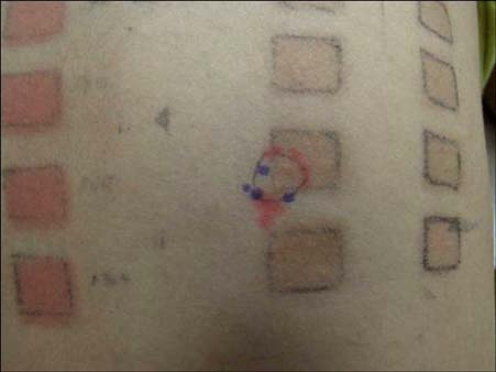
Fig. 5 The duplicate ultraviolet-A-irradiated sites demonstrate vesiculation.
Reference
-
1. Hann SK, Im S, Park YK, Lee S. Hydroa vacciniforme with unusually severe scar formation: diagnosis by repetitive UVA phototesting. J Am Acad Dermatol. 1991. 25:401–403.
Article2. Choi JH, Hann SK, Yoon MS, Choi BM, Ahn SK, Park YK. Hydroa vacciniforme: recurrence at adulthood and confirmative diagnosis by repetitive ultraviolet-A phototesting. Ann Dermatol. 1989. 1:83–86.3. Sonnex TS, Hawk JL. Hydroa vacciniforme: a review of ten cases. Br J Dermatol. 1988. 118:101–108.
Article4. Hawk JLM, Ferguson J, Hönigsmann H. Wolff K, Goldsmith LA, Katz SI, Gilchrest BA, Paller AS, Leffell DJ, editors. Hydroa vacciniforme. Fitzpatrick's dermatology in general medicine. 2008. 7th ed. New York: McGraw-Hill;820.5. Eramo LR, Garden JM, Esterly NB. Hydroa vacciniforme. Diagnosis by repetitive ultraviolet-A phototesting. Arch Dermatol. 1986. 122:1310–1313.
Article6. Epstein JH. Polymorphous light eruption. J Am Acad Dermatol. 1980. 3:329–343.
Article7. Elpern DJ, Morison WL, Hood AF. Papulovesicular light eruption. A defined subset of polymorphous light eruption. Arch Dermatol. 1985. 121:1286–1288.
Article8. Redeker AG, Bronow RS. Erythropoietic protoporphyria presenting as hydroa aestivale. Arch Dermatol. 1964. 89:104–109.
Article9. Wheeler CE, Cawley EP, Whitmore CW. Hydroa aestivale in identical twins. Arch Dermatol. 1960. 82:590–594.
Article10. Goldgeier MH, Nordlund JJ, Lucky AW, Sibrack LA, McCarthy MJ, McGuire J. Hydroa vacciniforme: diagnosis and therapy. Arch Dermatol. 1982. 118:588–591.
Article11. Cho KH, Li KS, Kim YK, Jeon YK, Kim CW, Lee SK, et al. Epstein-Barr virus associated lymphoproliferative lesion presenting as a hydroa vacciniforme-like eruption. Korean J Dermatol. 2004. 42:846–855.
