Ann Dermatol.
2014 Dec;26(6):773-774. 10.5021/ad.2014.26.6.773.
A Case of Sarcoidosis Presenting as Livedo
- Affiliations
-
- 1Department of Dermatology, Tokyo Medical and Dental University, Graduate School of Medicine, Tokyo, Japan. k.igawa.derm@tmd.ac.jp
- KMID: 2264883
- DOI: http://doi.org/10.5021/ad.2014.26.6.773
Abstract
- No abstract available.
MeSH Terms
Figure
-
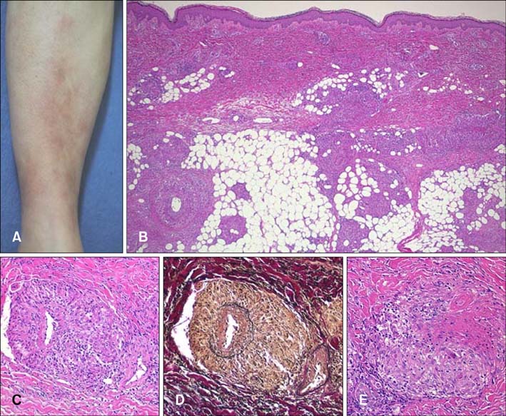
Fig. 1 (A) Erythema with subcutaneous nodules and livedo reticularis of the lower extremities. (B) Nodules of noncaseating epithelioid granulomas and diffuse dense mixed infiltrate of lymphocytes in the middle and lower dermis, and in subcutaneous tissue (H&E, ×20). (C) Granulomas surrounding damaged and narrowed blood vessels (H&E, ×100). (D) Granulomas surrounding damaged and narrowed blood vessels (Elastica van Gieson stain, ×100). (E) Some vessels were occluded with fibrin (H&E, ×100).
Reference
-
1. Haimovic A, Sanchez M, Judson MA, Prystowsky S. Sarcoidosis: a comprehensive review and update for the dermatologist: part I. Cutaneous disease. J Am Acad Dermatol. 2012; 66:699.e1–699.e18.2. Hayashi S, Hatamochi A, Hamasaki Y, Kitamura Y, Ishii Y, Fukuda T, et al. A case of sarcoidosis with livedo. Int J Dermatol. 2009; 48:1217–1221.
Article3. Takenoshita H, Yamamoto T. Erythema nodosum-like cutaneous lesions of sarcoidosis showing livedoid changes in a patient with sarcoidosis and Sjögren's syndrome. Eur J Dermatol. 2010; 20:640–641.4. Wei CH, Huang YH, Shih YC, Tseng FW, Yang CH. Sarcoidosis with cutaneous granulomatous vasculitis. Australas J Dermatol. 2010; 51:198–201.
Article5. Kawakami T, Soma Y. Successful use of mizoribine in a patient with sarcoidosis and cutaneous vasculitis. Acta Derm Venereol. 2011; 91:582–583.
Article
- Full Text Links
-
- Actions
-
Cited
- CITED
-
- Close
- Share
-
- Similar articles
-
- A Case of Livedo Reticularis Associated with Decompression Sickness
- Systemic Sarcoidosis Presenting with Arrhythmia
- A Case of Systemic Lupus Erythematosus and Secondary AntiphospholipidSyndrome Presenting as Livedo Reticularis
- A Case of Secondary Antiphospholipid Syndrome with Systemic Erythematosus Lupus Who Presenting Livedo Reticularis, Livedoid Vasculopathy, Peripheral Gangrene, and Leg Ulcers
- Scar Sarcoidosis after Blepharoplasty: A Case Series
