Ann Dermatol.
2014 Dec;26(6):771-773. 10.5021/ad.2014.26.6.771.
Sacrococcygeal Nodule in a Young Male Patient
- Affiliations
-
- 1Department of Dermatology, Ajou University School of Medicine, Suwon, Korea. hykang@ajou.ac.kr
- KMID: 2264882
- DOI: http://doi.org/10.5021/ad.2014.26.6.771
Abstract
- No abstract available.
Figure
-

Fig. 1 (A) Solitary skin-colored nodule with scales is located in coccyx. (B) Hyperkeratosis and upperdermal edema, and marked increased dermal thickness are observed at the lower magnification (H&E, ×10). (C) Thickening of individual collagen bundles are noted at the higher magnification (H&E, ×100).
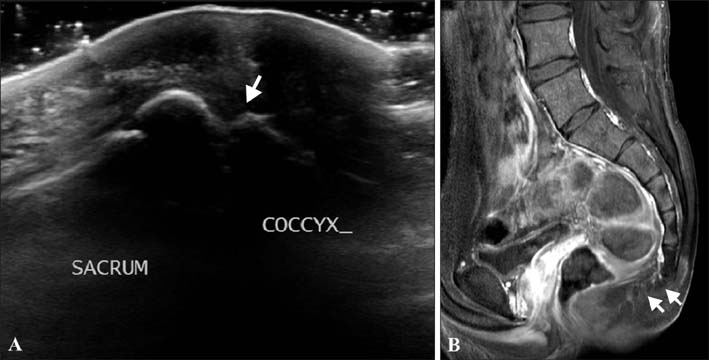
Fig. 2 (A) Abrupt angle of sacrococcygeal junction (arrow) under the nodular skin lesion was visualizedthroughsonogram. (B) Anterior dislocation of the coccyx (arrows) was visualized through T1-weighted sagittal view ofmagnetic resonance imaging examination.
Reference
-
1. Nakamura A, Inoue Y, Ishihara T, Matsunaga W, Ono T. Acquired coccygeal nodule due to repeated stimulation by a bicycle saddle. J Dermatol. 1995; 22:365–369.
Article2. Hashimoto I, Shono Y, Ishida S, Nakanishi H. Developmental mechanism of juvenile coccygeal fibrosis (so-called coccygeal pad). J Dermatol. 2013; 40:832–836.
Article3. Dekio I, Murata T. Coccygeal pad. Contact Dermatitis. 2003; 48:234–235.
Article4. Mullen M, Rabban J, Frieden IJ. Sacrococcygeal teratoma masquerading as congenital hemangioma. Pediatr Dermatol. 2013; 30:112–116.
Article5. de Parades V, Bouchard D, Janier M, Berger A. Pilonidal sinus disease. J Visc Surg. 2013; 150:237–247.
Article
