Rare multiple variations in brachial plexus and related structures in the left upper limb of a Dravidian male cadaver
- Affiliations
-
- 1Department of Anatomy, Velammal Medical College Hospital and Research Institute, Madurai, India. davidebenezer@yahoo.com
- 2Department of Anatomy, All India Institute of Medical Sciences, Bhopal, India.
- KMID: 2263110
- DOI: http://doi.org/10.5115/acb.2013.46.2.163
Abstract
- Anatomical variations of the nerves, muscles, and vessels in the upper limb have been described in many anatomical studies; however, the occurrence of 6 variations in an ipsilateral limb is very rare. These variations occur in the following structures: the pectoralis minimus muscle, the communication between the external jugular vein and cephalic vein, axillary arch, the Struthers ligament, the medial, lateral, and posterior cords of the brachial plexus, and the common arterial trunk from the third part of the axillary artery. The relationship of these variations to each other and their probable clinical presentation is discussed.
Keyword
MeSH Terms
Figure
-
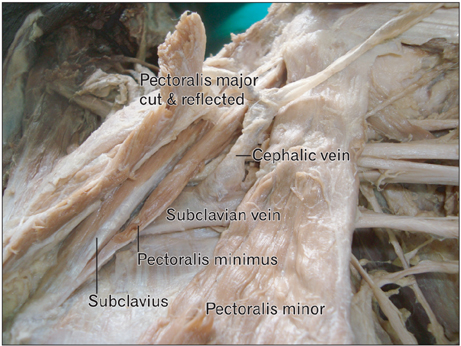
Fig. 1 Left pectoral region showing the cephalic vein separating the pectoralis minimus from the pectoralis minor muscle.
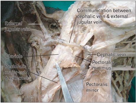
Fig. 2 Left pectoral region showing the communication of the cephalic vein with the external jugular vein, which was located superficial to the clavicle.
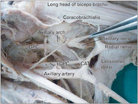
Fig. 3 Left axilla showing the axillary arch and common arterial trunk (CAT) from the axillary artery, which bifurcated the posterior cord. LDPC, lower division of the posterior cord; UDPC, upper division of the posterior cord.

Fig. 4 Anterior view of the left upper arm showing variations in the medial and lateral cords. AA, axillary artery; BA, brachial artery; BV, basilic vein; LC, lateral cord; LD, lateral division of the lateral cord; LRM, lateral root of the median nerve; MC, medial cord; MCN, musculocutaneous nerve; MD, medial division of the lateral cord; MN, median nerve; MRM, medial root of the median nerve; NCB, nerve to the coracobrachialis; UN, ulnar nerve.
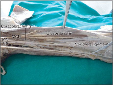
Fig. 5 Anterior view of the left arm, showing the median nerve (MN) and the brachial artery (BA) running deep to the Struthers ligament, with muscle fibers arising from it. MCN, musculocutaneous nerve.
Cited by 3 articles
-
Bilateral absence of subclavius muscles with thickened costocoracoid ligaments: a case report with the clinical-anatomical correlation
Kasapuram Dheeraj, Harisha K. Sudheer, Subhash Bhukiya, Neerja Rani, Seema Singh
Anat Cell Biol. 2022;55(2):255-258. doi: 10.5115/acb.21.246.Uncommon configuration of intercostobrachial nerves, lateral roots, and absent medial cutaneous nerve of arm in a cadaveric study
Rosemol Xaviour
Anat Cell Biol. 2023;56(4):570-574. doi: 10.5115/acb.23.149.Anatomical variations and surgical implications of axillary artery branches: an anatomical study of the coracoid process region
Pawaree Nonthasaen, Thawanthorn Chaimongkhol, Thanapon Chobpenthai, Pasuk Mahakkanukrauh
Anat Cell Biol. 2025;58(1):35-43. doi: 10.5115/acb.24.215.
Reference
-
1. Standring S. Gray's anatomy: the anatomical basis of clinical practice. 2005. 39th ed. London: Elsevier Churchill Livingstone.2. Nayak BS, Soumya KV. Abnormal formation and communication of external jugular vein. Int J Anat Var. 2008. 1:15–16.3. Bakirci S, Kafa IM, Uysal M, Sendemir E. Langer's axillary arch (axillopectoral muscle): a variation of latissimus dorsi muscle. Int J Anat Var. 2010. 3:91–92.4. Pai MM, Rajanigandha , Prabhu LV, Shetty P, Narayana K. Axillary arch (of Langer): incidence, innervation, importance. Online J Health Allied Sci. 2006. 5:4.5. Bertha A, Kulkarni NV, Maria A, Jestin O, Joseph K. Entrapment of deep axillary arch by two roots of radial nerve: An anatomical variation. J Anat Soc India. 2009. 58:40–43.6. Iamsaard S, Uabundit N, Khamanarong K, Sripanidkulchai K, Chaiciwamongkol K, Namking M, Ratanasuwan S, Boonruangsri P, Hipkaeo W. Duplicated axillary arch muscles arising from the latissimus dorsi. Anat Cell Biol. 2012. 45:288–290.7. Pillay M, Jacob SM. Bilateral presence of axillary arch muscle passing through the posterior cord of the brachial plexus. Int J Morphol. 2009. 27:1047–1050.8. Guy MS, Sandhu SK, Gowdy JM, Cartier CC, Adams JH. MRI of the axillary arch muscle: prevalence, anatomic relations, and potential consequences. AJR Am J Roentgenol. 2011. 196:W52–W57.9. Bhat KM, Gowda S, Potu BK. Nerve loop around the axillary vessels by the roots of the median nerve a rare variation in a south Indian male cadaver: a case report. Cases J. 2009. 2:179.10. Siqueira MG, Martins RS. The controversial arcade of Struthers. Surg Neurol. 2005. 64:Suppl 1. S1:17–S1:20.
- Full Text Links
-
- Actions
-
Cited
- CITED
-
- Close
- Share
-
- Similar articles
-
- Multiple unilateral variations in medial and lateral cords of brachial plexus and their branches
- Bilateral absence of musculocutaneous nerve with unusual branching pattern of lateral cord and median nerve of brachial plexus
- Anatomical variation of median nerve: cadaveric study in brachial plexus
- Bilateral single cord of the brachial plexus in an adult female cadaver of South Indian origin
- Uncommon configuration of intercostobrachial nerves, lateral roots, and absent medial cutaneous nerve of arm in a cadaveric study
