Congenital Horner Syndrome with Heterochromia Iridis Associated with Ipsilateral Internal Carotid Artery Hypoplasia
- Affiliations
-
- 1Department of Radiology, Cliniques Universitaires Saint-Luc, UCL, Brussels, Belgium. fabrice.deprez@uclouvain.be
- 2Department of Ophthalmology, Cliniques Universitaires Saint-Luc, UCL, Brussels, Belgium.
- KMID: 2242666
- DOI: http://doi.org/10.3988/jcn.2015.11.2.192
Abstract
- BACKGROUND
Horner syndrome (HS), also known as Claude-Bernard-Horner syndrome or oculosympathetic palsy, comprises ipsilateral ptosis, miosis, and facial anhidrosis.
CASE REPORT
We report herein the case of a 67-year-old man who presented with congenital HS associated with ipsilateral hypoplasia of the internal carotid artery (ICA), as revealed by heterochromia iridis and confirmed by computed tomography (CT).
CONCLUSIONS
CT evaluation of the skull base is essential to establish this diagnosis and distinguish aplasia from agenesis/hypoplasia (by the absence or hypoplasia of the carotid canal) or from acquired ICA obstruction as demonstrated by angiographic CT.
Keyword
MeSH Terms
Figure
-
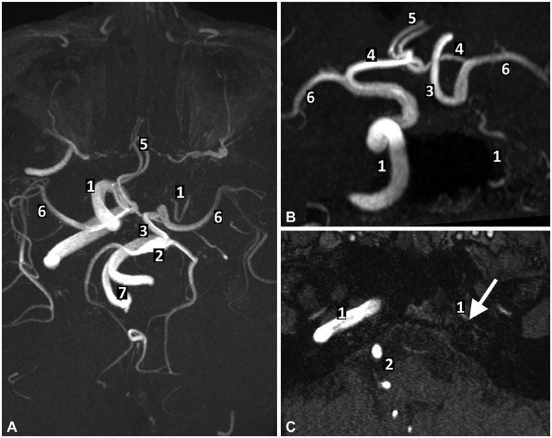
Fig. 1 Magnetic resonance imaging series. Axial (A) and coronal (B) maximal intensity projections of three-dimensional time-of-flight (TOF) angiographic series demonstrating an extremely spindly left internal carotid artery (ICA). The left anterior cerebral artery (ACA) and middle cerebral artery (MCA) are supplied both by a permeable anterior communicating artery and a large posterior communicant artery (PCoA) from the basilar artery (BA). (C) Axial TOF angiographic study showing an important narrowing of the left ICA (white arrow). 1: ICA, 2: BA, 3: left PCoA, 4: ACA segment A1, 5: ACA segment A2, 6: MCA, 7: vertebral arteries.
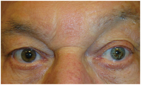
Fig. 2 Heterochromia with left-iris hypopigmentation and left-sided miosis. This patient compensates for a left superior palpebral ptosis by active raising his left eyebrow.
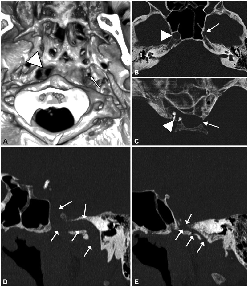
Fig. 3 Unenhanced skull CT. 3D volume-rendering reconstruction of (A) the inferior face of the skull base and (B) axial view of the skull base, demonstrating a right normal carotid canal (white arrowhead) and a left highly hypoplastic carotid canal (white arrow). On axial maximal intensity projection (MIP) view of the cavernous and clinoid segment of the internal carotid artery (ICA) (C4-C5) and the sella turcica (C), it can be seen that the left hypoplastic ICA is not affected by atheromatous calcifications. MIP sagittal oblique reconstructions of the carotid canals (white arrows) demonstrate a right normal carotid canal (D) and a left highly hypoplastic carotid canal (E).
Reference
-
1. Ibrahim M, Branson HM, Buncic JR, Shroff MM. A case of Horner syndrome with intermittent mydriasis in a patient with hypoplasia of the internal carotid artery. AJNR Am J Neuroradiol. 2006; 27:1318–1320.2. Jeffery AR, Ellis FJ, Repka MX, Buncic JR. Pediatric Horner syndrome. J AAPOS. 1998; 2:159–167.
Article3. Given CA 2nd, Huang-Hellinger F, Baker MD, Chepuri NB, Morris PP. Congenital absence of the internal carotid artery: case reports and review of the collateral circulation. AJNR Am J Neuroradiol. 2001; 22:1953–1959.4. Lie TA. Excerpta Medica Foundation. Congenital anomalies of the carotid arteries: including the carotid-basilar and carotid-vertebral anastomoses; an angiographic study and a review of the literature. Amsterdam: Excerpta Medica Foundation;1968.5. Farhat W, Ahdab R, Hosseini H. Congenital agenesis of internal carotid artery with ipsilateral Horner presenting as focal neurological symptoms. Vasc Health Risk Manag. 2011; 7:37–40.6. Hüttemann K, Nowe T, Engelhorn T, Doerfler A, Schellinger PD, Köhrmann M. Ischemic stroke and subarachnoid hemorrhage, two sides of a story: congenital absence of the internal carotid artery. Two cases and review of the literature. J Neurol. 2008; 255:1985–1987.
Article7. Florio F, Balzano S, Nardella M, Strizzi V, Cammisa M, Bozzini V, et al. Congenital absence of the internal carotid artery. Cardiovasc Intervent Radiol. 1999; 22:74–78.
Article8. Ryan FH, Kline LB, Gomez C. Congenital Horner's syndrome resulting from agenesis of the internal carotid artery. Ophthalmology. 2000; 107:185–188.
Article9. Imesch PD, Wallow IH, Albert DM. The color of the human eye: a review of morphologic correlates and of some conditions that affect iridial pigmentation. Surv Ophthalmol. 1997; 41:Suppl 2. S117–S123.
Article10. Gladstone RM. Development and significance of heterochromia of the iris. Arch Neurol. 1969; 21:184–191.
Article
- Full Text Links
-
- Actions
-
Cited
- CITED
-
- Close
- Share
-
- Similar articles
-
- A Case of Pericarotid Syndrome Associated with Malignant Lymphoma
- Congenital External Carotid-Internal Carotid Artery Anastomosis
- Painful Horner syndrome after thyroidectomy: A case report
- Congenital Hypoplasia of the Bilateral Internal Carotid Artery: A Case Report
- A Case of Ocular Ischemic Syndrome Associated with Internal Carotid Artery Obstruction with Atheroma Diagnosed by Doppler Ultrasonography
