Imaging Sci Dent.
2013 Dec;43(4):289-293. 10.5624/isd.2013.43.4.289.
Image analysis of the eruptive positions of third molars and adjacent second molars as indicators of age evaluation in Thai patients
- Affiliations
-
- 1Department of Oral Biology and Diagnostic Sciences and Biomedical Engineering Centre, Faculty of Dentistry, Chiang Mai University, Chiang Mai, Thailand. drmaymh@gmail.com
- 2Department of Computer Engineering and Biomedical Engineering Centre, Faculty of Engineering, Chiang Mai University, Chiang Mai, Thailand.
- KMID: 2229616
- DOI: http://doi.org/10.5624/isd.2013.43.4.289
Abstract
- PURPOSE
This study was performed to determine the relationship between the stage of tooth eruption (both vertical and mesio-angular) and chronological age.
MATERIALS AND METHODS
Indirect digital panoramic radiographs were used to measure the distances from the dentinoenamel junction (DEJ) of the second molars to the occlusal plane of the second molar teeth and of the adjacent third molars in 264 Thai males and 437 Thai females using ImageJ software. The ratio of those distances was calculated by patient age, and the correlation coefficient of the ratio of the third molar length to the second molar length was calculated.
RESULTS
The correlation between the height of the vertically erupted upper third molar teeth and age was at the intermediate level. The age range of > or =15 to <16 years was noted to be the range in which the correlation between the chronological age determined from the eruptional height and actual chronological age was statistically significant. The mean age of the female subjects, in which the position of the right upper third molar teeth was at or above the DEJ of the adjacent second molar but below one half of its coronal height was 19.9+/-2.6 years. That for the left side was 20.2+/-2.7 years. The mean ages of the male subjects were 20.1+/-3.3 years and 19.8+/-2.7 years for the right and left sides, respectively.
CONCLUSION
It might be possible to predict chronological age from the eruption height of the wisdom teeth.
MeSH Terms
Figure
-
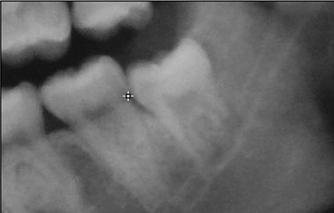
Fig. 1 Cropped image of 350×250 pixels using ImageJ software. The reference point (marker) is located at the dentino-enamel junction.
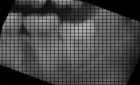
Fig. 2 A rotated radiograph achieved the paralleling of the second molar's occlusal plane to one of the grid lines.
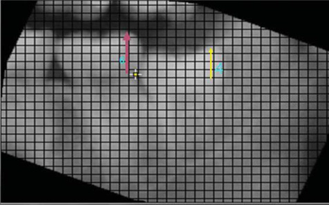
Fig. 3 Measurement from the reference point to the highest point of the second molar and third molar occlusal plane. The score 6 is marked for the second molar and the score 4 is marked for the third molar.
Cited by 1 articles
-
Accuracy of an equation for estimating age from mandibular third molar development in a Thai population
Karune Verochana, Sangsom Prapayasatok, Apirum Janhom, Phattaranant May Mahasantipiya, Narumanas Korwanich
Imaging Sci Dent. 2016;46(1):1-7. doi: 10.5624/isd.2016.46.1.1.
Reference
-
1. Levesque GY, Demirijian A, Tanguay R. Sexual dimorphism in the development, emergence, and agenesis of the mandibular third molar. J Dent Res. 1981; 60:1735–1741.
Article2. Olze A, Peschke C, Schulz R, Schmeling A. Studies of the chronological course of wisdom tooth eruption in a German population. J Forensic Leg Med. 2008; 15:426–429.
Article3. Olze A, van Niekerk P, Ishikawa T, Zhu BL, Schulz R, Maeda H, et al. Comparative study on the effect of ethnicity on wisdom tooth eruption. Int J Legal Med. 2007; 121:445–448.
Article4. Uzamis M, Kansu O, Taner TU, Alpar R. Radiographic evaluation of third-molar development in a group of Turkish children. ASDC J Dent Child. 2000; 67:136–141.5. Sarnat H, Kaffe I, Porat J, Amir E. Developmental stages of the third molar in Israeli children. Pediatr Dent. 2003; 25:373–377.6. Blankenship JA, Mincer HH, Anderson KM, Woods MA, Burton EL. Third molar development in the estimation of chronologic age in American blacks as compared with whites. J Forensic Sci. 2007; 52:428–433.
Article7. Meinl A, Tangl S, Huber C, Maurer B, Watzek G. The chronology of third molar mineralization in the Austrian population-a contribution to forensic age estimation. Forensic Sci Int. 2007; 169:161–167.
Article8. Mincer HH, Harris EF, Berryman HE. The A.B.F.O. study of third molar development and its use as an estimator of chronological age. J Forensic Sci. 1993; 38:379–390.
Article9. Nykanen R, Espeland L, Kvaal SI, Krogstad O. Validity of the Demirjian method for dental age estimation when applied to Norwegian children. Acta Odontol Scand. 1998; 56:238–244.10. Orhan K, Ozer L, Orhan AI, Dogan S, Paksoy CS. Radiographic evaluation of third molar development in relation to chronological age among Turkish children and youth. Forensic Sci Int. 2007; 165:46–51.
Article11. Dhanjal KS, Bhardwaj MK, Liversidge HM. Reproducibility of radiographic stage assessment of third molars. Forensic Sci Int. 2006; 159:Suppl 1. s74–s77.
Article12. Demirjian A, Goldstein H, Tanner JM. A new system of dental age assessment. Hum Biol. 1973; 45:211–227.13. Thevissen PW, Pittayapat P, Fieuws S. Estimating age of majority on third molars developmental stages in young adults from Thailand using a modified scoring technique. J Forensic Sci. 2009; 54:428–432.
Article
- Full Text Links
-
- Actions
-
Cited
- CITED
-
- Close
- Share
-
- Similar articles
-
- Prevalence of missing and impacted third molars in adults aged 25 years and above
- Third molar changes following lower second molar extractions
- Analysis of the Characteristics of First Permanent Molars with Delayed Eruption
- A study of the calcification of the second and the third molars in skeletal Class II and III malocclusions
- Analysis of Prevalence of Pyramidal Molars in Adolescent
