A Huge Mediastinal Organizing Hematoma Causing Reversal of Atrial Septal Defect Shunt Flow
- Affiliations
-
- 1Division of Cardiology, Department of Internal Medicine, Samsung Medical Center, Sungkyunkwan University School of Medicine, Seoul, Korea. sclee@skku.edu
- 2Department of Radiology and Center of Imaging Science, Cardiovascular Imaging Center, Samsung Medical Center, Sungkyunkwan University School of Medicine, Seoul, Korea.
- 3Department of Thoracic and Cardiovascular Surgery, Samsung Medical Center, Sungkyunkwan University School of Medicine, Seoul, Korea.
- KMID: 2225142
- DOI: http://doi.org/10.4070/kcj.2011.41.2.97
Abstract
- We report a case of a 46-year-old woman who presented with subacute exertional dyspnea and severe hypoxia. A large cystic mass compressing the right side of the heart along with right-to-left atrial shunt flow through an alleged atrial septal defect (ASD) were detected on echocardiography. CT scan of the chest and MRI of the heart revealed a loculated cystic mediastinal mass with hemorrhage measuring 5.5x8 cm compressing the right atrium and ventricle. The patient underwent cyst resection and primary closure of the ASD. This report illustrates a case of an unusual symptomatic pericardial mass compressing the right atrium and ventricle in a patient with an secundum ASD.
Keyword
MeSH Terms
Figure
-
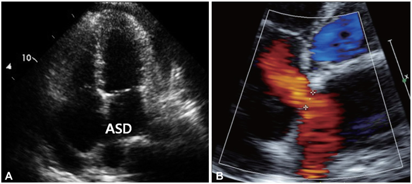
Fig. 1 Initial echocardiogram. Apical four-chamber view is showing an atrial septal defect (A and B). On color doppler echocardiogram, the direction of the shunt flow through the septal defect is shown to be left-to-right.
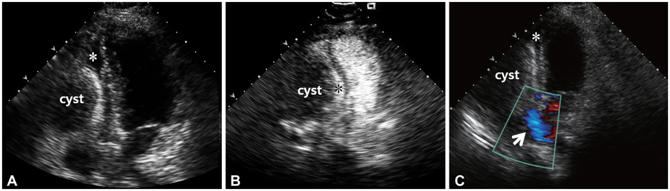
Fig. 2 Follow-up echocardiogram. A: in apical four-chamber view, the pericardial cyst is compressing the right side of the heart (*) almost completely. B: contrast echocardiogram demonstrating the pericardial cyst compressing the right atrium and ventricle (*). Cystic mass does not show enhancement of contrast. C: color doppler echocardiogram showing a huge pericardial cyst and an atrial septal defect. The direction of the shunt flow (white arrow) through the septal defect is shown to be right-to-left.
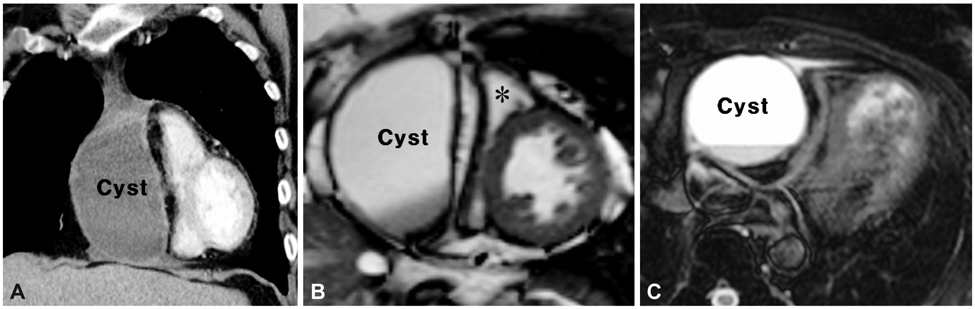
Fig. 3 Chest CT and MRI of the heart. Coronal view of chest CT (A) shows a cystic lesion in the right pericardial space. A short axis cine image view (B) demonstrates a compressed right ventricle (*) and an intact left ventricle. There is no communication between the mass and the heart. Axial T2-weighted view (C) shows a dark fluid-fluid level within the dependent portion of the mass.
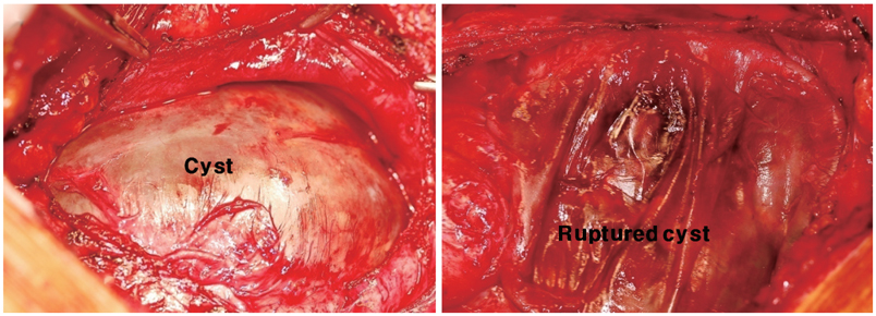
Fig. 4 Intra-operative photograph of the pericardial cyst (patient's head is on the left side).
Reference
-
1. Koshy T, Sinha PK, Misra S, Unnikrishnan M. Pericardial cyst. Ann Card Anaesth. 2008. 11:129–130.2. Modi S, Chenzbraun A, Fewins H, Binukrishnan S, Ramsdale DR. Giant asymptomatic pericardial cyst. J Cardiovasc Med (Hagerstown). 2009. 10:646–648.3. Borgna-Pignatti C, Andreis IB, Rugolotto S, Balter R, Bontempini L. Thymic cyst appearing after treatment of mediastinal non-Hodgkin lymphoma. Med Pediatr Oncol. 1994. 22:70–72.4. Elamin WF, Hannan K. Pericardial cyst: an unusual cause of pneumonia. Cases J. 2008. 1:26.5. Tanoue Y, Fujita S, Kanaya Y, Tominaga R. Acute cardiac tamponade due to a bleeding pericardial cyst in a 3-year-old child. Ann Thorac Surg. 2007. 84:282–284.6. Borges AC, Gellert K, Dietel M, Baumann G, Witt C. Acute right-sided heart failure due to hemorrhage into a pericardial cyst. Ann Thorac Surg. 1997. 63:845–847.7. Bandeira FC, de Sa VP, Moriguti JC, et al. Cardiac tamponade: an unusual complication of pericardial cyst. J Am Soc Echocardiogr. 1996. 9:108–112.8. Ogah OS, Akisanya CO, Timeyin AO, Adebiyi AA, Adebo OA. A large pericardial cyst presenting with severe chest pain: a case report and review of literature. Afr J Med Med Sci. 2009. 38:83–86.9. Chopra PS, Duke DJ, Pellett JR, Rahko PS. Pericardial cyst with partial erosion of the right ventricular wall. Ann Thorac Surg. 1991. 51:840–841.10. Lesniak-Sobelga AM, Olszowska M, Tracz W, et al. Giant pericardial cyst compressing the right ventricle. Ann Thorac Surg. 2008. 85:1811.11. Komodromos T, Lieb D, Baraboutis J. Unusual presentation of a pericardial cyst. Heart Vessels. 2004. 19:49–51.12. Lee JA, Kim BS, Cho HJ, et al. A case of constrictive pericarditis with localized pericardial effusion simulating a cystic mass. Korean Circ J. 1991. 21:791–796.13. Jin KH, Lee WS, Lee IK, Kim KS, Kim YN, Kim KB. Pericardial cysts: three cases report. Korean Circ J. 1987. 17:795–801.14. Brickner ME, Hillis LD, Lange RA. Congenital heart disease in adults: second of two parts. N Engl J Med. 2000. 342:334–342.
- Full Text Links
-
- Actions
-
Cited
- CITED
-
- Close
- Share
-
- Similar articles
-
- A Case of Atrial Septal Defect in Identical Twins
- Echocardiographic Examination of the Right Pulmonary Artery in the Patients with Left to Right Shunt
- Atrial Septal Defect with Normal Pulmonary Arterial Pressure in Adult Cyanotic Patient
- A Case of Atrial Septal Aneurysm Associated with Atrial Septal Defect
- The Significance of a Crochetage Pattern on R Wave in Electrocardiographic Inferior Limb Leads in Atrial Septal Defect
