Supraventricular Tachycardia and Sinus Rhythm with Contralateral Bundle Branch Block Patterns
- Affiliations
-
- 1Krannert Institute of Cardiology, Indiana University School of Medicine, Indianapolis, IN, USA. midas@iupui.edu
- KMID: 2223890
- DOI: http://doi.org/10.4070/kcj.2014.44.4.271
Abstract
- A contralateral bundle branch block (BBB) aberration during tachycardia with a preexisting BBB strongly suggests the presence of ventricular tachycardia. We report on a middle-aged, female patient presented with wide QRS tachycardia. The patient had orthodromic atrioventricular tachycardia with a left BBB aberration in the presence of a preexisting right BBB due to an abnormal His-Purkinje system. We learned that the contralateral BBB aberration with supraventricular tachycardia could be seen when the His-Purkinje system was abnormal.
MeSH Terms
Figure
-

Fig. 1 12-lead surface electrocardiograms. (A) shows the baseline normal sinus rhythm. (B) shows sinus rhythm with right bundle branch block after mechanical trauma. (C) shows a left bundle branch block morphology tachycardia with a rate of 200 beats/min.
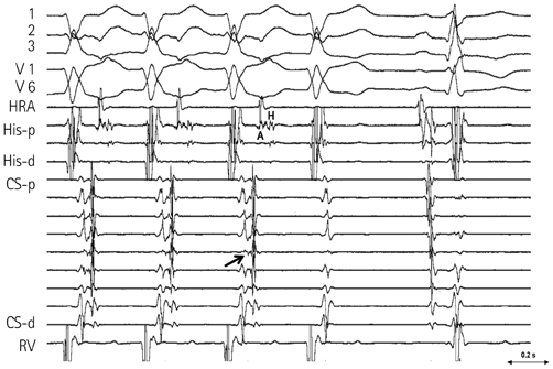
Fig. 2 The surface electrocardiogram and the intracardiac electrograms during the tachycardia. Tachycardia terminated spontaneously without atrial activation. The earliest retrograde atrial (A) activation site was in the mid-CS (arrow). The His (H) activation followed the atrial activation and occurred before the beginning of the QRS. p: proximal, d: distal, CS: coronary sinus, HRA: high right atrium, RV: right ventricle.

Fig. 3 Right ventricular burst pacing with a cycle length of 420 msec after AP ablation. His potential was obtained from left ventricle. During RV pacing, there was progressive prolongation of VH interval (arrows) that ended up with VA conduction block (Wenckebach VA block at below the His). p: proximal, d: distal, HRA: high right atrium, CS: coronary sinus, AP: accessory pathway, RV: right ventricle, VH: ventriculo-Hisian, VA: ventriculo-atrial.

Fig. 4 Burst pacing and programmed stimulation from HRA. The top panel shows that burst pacing from the HRA at 280 msec prolonged the interval between the His and RB, and resulted in intra/infra Hisian block (big arrow; Wenckebach block). The lower 3 panels show programmed single extra-stimulation with a drive cycle length of 500 msec. With extrastimuli at 400 msec, the QRS morphology continued to exhibit right bundle branch block. As the coupling interval was shortened, the QRS developed left bundle branch block morphology after prolongation of the intra/infra Hisian conduction (small arrows) and the RB to V interval became longer and fixed. p: proximal, d: distal, HRA: high right atrium, AH: A to H interval, HR: H to RB interval, RV: RB to V interval, @: at, scale: msec.
Reference
-
1. Approach to wide QRS tachycardias. In : Issa Z, Miller J, Zipes D, editors. Clinical arrhythmology and electrophysiology. 1st ed. Philadelphia: WB Saunders;2009. p. 398.2. de P.dua F, Pereirinha A, Marques N, Lopes MG, Macfarlane PW. Chapter 14. Conduction defect. In : Macfarlane PW, Oosterom A, Pahlm O, Kligfield P, Janse M, Camm J, editors. Comprehensive Electrocardiology. 2nd ed. London: Springer;2011. p. 554.3. Demoulin JC, Kulbertus HE. Histopathological examination of concept of left hemiblock. Br Heart J. 1972; 34:807–814.
- Full Text Links
-
- Actions
-
Cited
- CITED
-
- Close
- Share
-
- Similar articles
-
- Preexcitation Syndrome with a Mahaim-type Accessory Pathway
- A Case of Hyperthyroidism with Complete Atrioventricular Block and Cardiac Arrest
- Differential Diagnosis of Supraventricular Tachycardia
- Rate-dependent Left Bundle Branch Block during General Anesthesia : A case report
- Studies on Electrocardiogram of 18,000 Koreans
