A Case of Kikuchi Disease Accompanied with Bilateral Retinal Vasculitis
- Affiliations
-
- 1Department of Internal Medicine, Jeju National University School of Medicine, Jeju, Korea. slera@jejunuh.ac.kr
- 2Department of Ophthalmology, Jeju National University School of Medicine, Jeju, Korea.
- 3Department of Pathology, Jeju National University School of Medicine, Jeju, Korea.
- KMID: 2223155
- DOI: http://doi.org/10.4078/jrd.2011.18.3.220
Abstract
- Kikuchi disease, also called histiocytic necrotizing lymphadenitis, is an uncommon, idiopathic and generally self-limited disease, characterized by cervical lymphadenopathy. It can present systemic symptoms and signs, but ocular involvement is unusual. We report a 35-year-old woman who presented sudden decreased visual acuity and a swollen lymph node on the left side of her neck. On laboratory findings, there were no evidences of infection, autoimmune disease and systemic vasculitis. She was diagnosed with Kikuchi disease and bilateral retinal vasculitis by histologic analysis of lymph node, fundoscopy and fluorescein angiography.
MeSH Terms
Figure
-
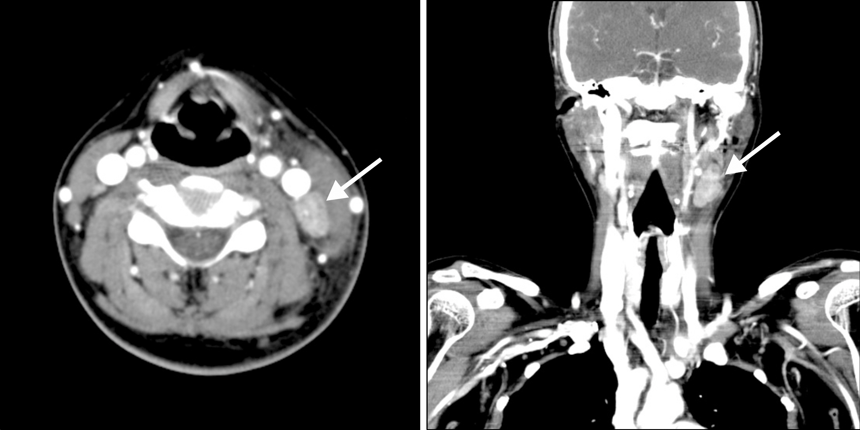
Figure 1. Neck CT: There was a 3.3×1.8 cm sized, partially necrotized cervical lymph node from the inside of the left upper sternocl-eidomastoid (SCM) muscle (arrow). It was the biggest node and several other enlarged, but smaller lymph nodes were also found along the inside of the left SCM muscle
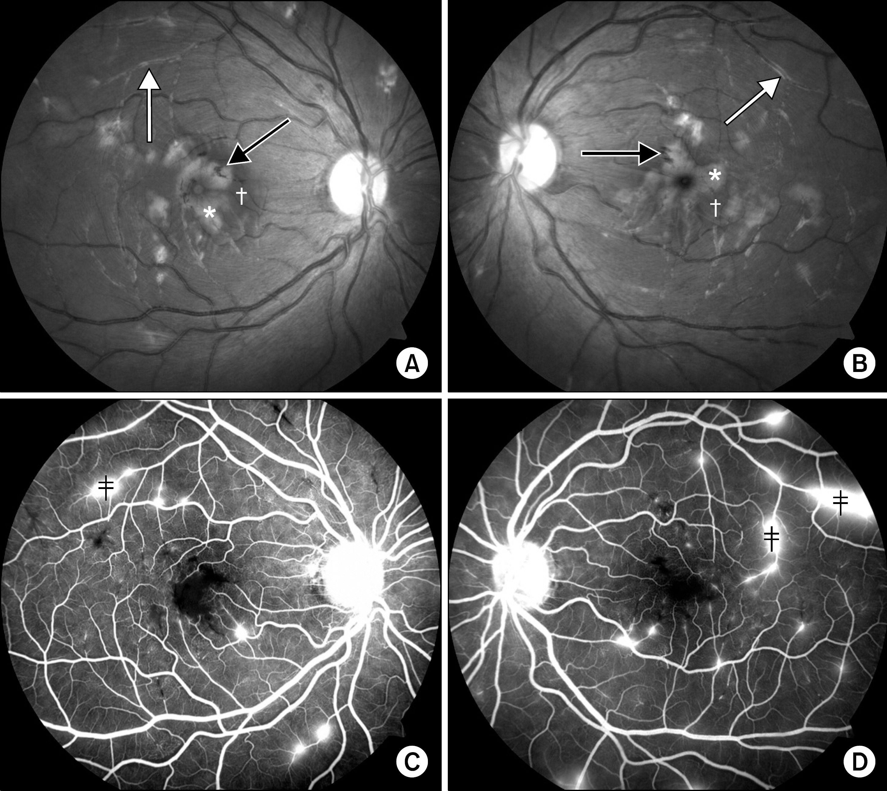
Figure 2. (A, B) Edema (†), soft exudates (∗) and flame shaped hemorrhage (black arrow) could be found around macula on fundos-copic examination. Also, there were several vascular sheathings (white arrow), the hallmark of retinal vasculitis. (C, D) On fluorescein angio-graphy, there were some vascular leakages (‡).

Figure 3. (A) On H & E stain, patches of necrosis (∗) and area of histiocyte proliferation (white arrow) were observed (100×). (B) Histiocytes were positive for CD68 (black arrow, 100× optical resolution).
Reference
-
References
1. Kikuchi M. Lymphadenitis showing focal reticulum cell hyperplasia with nuclear debris and phagocytes. Nippon Ketsueki Gakkai Zasshi. 1972; 35:379–80.2. Fujimoto Y, Kojima Y, Yamaguchi K. Cervical subacute necrotizing lymphadenitis. Naika. 1972; 30:920–7.3. Mohanty SK, Arora R, Saha M. Kikuchi-Fujimoto disease: an overview. J Dermatol. 2002; 29:10–4.
Article4. Zou W, Wen F. Bilateral occlusive retinal vasculitis in Kikuchi-Fujimoto disease. Clin Experiment Ophthalmol. 2007; 35:875–7.
Article5. Taguri AH, McIlwaine GG. Bilateral panuveitis: a possi-ble association with Kikuchi-Fujimoto disease. Am J Ophthalmol. 2001; 132:419–21.
Article6. Norris AH, Krasinskas AM, Salhany KE, Gluckman SJ. Kikuchi-Fujimoto disease: a benign cause of fever and lymphadenopathy. Am J Med. 1996; 101:401–5.
Article7. Dorfman RF, Berry GJ. Kikuchi's histiocytic necrotizing lymphadenitis: an analysis of 108 cases with emphasis on differential diagnosis. Semin Diagn Pathol. 1988; 5:329–45.8. Anagnostopoulos I, Hummel M, Korbjuhn P, Papadaki T, Anagnostou D, Stein H. Epstein-Barr virus in Kikuchi-Fujimoto disease. Lancet. 1993; 341:893.
Article9. Pileri SA, Facchetti F, Ascani S, Sabattini E, Poggi S, Piccioli M, et al. Myeloperoxidase expression by histiocytes in Kikuchi's and Kikuchi-like lymphadenopathy. Am J Pathol. 2001; 159:915–24.
Article10. Lee J, Lee IH, Kim TH, Jun JB, Jung SS, Ahn MJ, et al. A case of SLE associated with histiocytic necrotizing lymphadenitis. J Korean Rheum Assoc. 1998; 5:236–42.11. Kim SH, Kim SJ, Chung H, Lee HS, Kim HB, Park KH. Bilateral anterior uveitis as an unusual manifestation of Kikuchi-Fujimoto disease. Rheumatology (Oxford). 2004; 43:1056–7.
Article12. Rocher F, Pelosse B, Momtchilova M, Laroche L. Kikuchi's disease and ocular manifestation. J Fr Ophtalmol. 2006; 29:932–6.13. Pérez Alvarez MJ, Moreno López M. Panuveitis as a pos-sible ophthalmic complication of Kikuchi-Fujimoto disease. Arch Soc Esp Oftalmol. 2005; 80:41–4.14. Duker JS, Brown GC, Brooks L. Retinal vasculitis in Crohn's disease. Am J Ophthalmol. 1987; 103:664–8.
Article
- Full Text Links
-
- Actions
-
Cited
- CITED
-
- Close
- Share
-
- Similar articles
-
- A Case of Tuberculosis-related Retinal Vasculitis
- A Case of Acute Multifocal Hemorrhagic Retinal Vasculitis
- A Case of Juvenile Dermatomyositis Accompanied by Hemorrhagic Retinal Vasculitis and Multiple Gastrointestinal Bleeding
- A Case of Kikuchi Fujimoto's Disease Accompanied by Hemophagocytic Lymphohistiocytosis
- Bilateral Frosted Branch Angiitis in Kikuchi-Fujimoto Disease
