Dose-Related Effect of Extracorporeal Shock Wave Therapy for Plantar Fasciitis
- Affiliations
-
- 1Department of Rehabilitation Medicine, Gwangju Veterans Hospital, Gwangju, Korea. standupmd@hanmail.net
- KMID: 2219535
- DOI: http://doi.org/10.5535/arm.2013.37.3.379
Abstract
OBJECTIVE
To examine the dose-related effect of extracorporeal shock wave therapy (ESWT) for plantar fasciitis.
METHODS
Sixty patients with plantar fasciitis despite conservative treatment were enrolled. The patients were divided into a low-energy group (group L: n=30, 1,000 shocks/session, energy flux density [EFD] per shock 0.08 mJ/mm2) and a medium-energy group (group M: n=30, 1,000 shocks/session, EFD 0.16 mJ/mm2). The main outcome measures were visual analogue scale (VAS), Roles and Maudsley (RM) score, and thickness of plantar fascia (PF). To compare the effects between each group, follow-up was carried out 1 week after 3 and 6 sessions, and 1 and 3 months after ESWT.
RESULTS
Significant VAS and RM score improvement, and PF thickness reduction were observed in both groups (p<0.01). After 3 sessions of ESWT, group M showed significant improvement in the VAS and RM score than group L, whereas after 3 additional sessions applied in group L, the main outcomes were no longer significantly different in both groups (p>0.05).
CONCLUSION
Therapeutic effect might disclose a dose-related relationship; therefore, EFD and the times of the session are considerable factors when treating with ESWT.
Figure
-
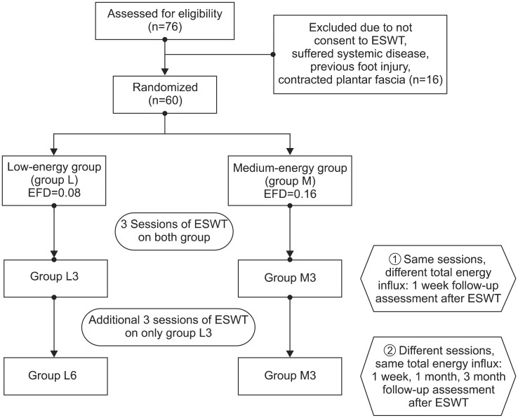
Fig. 1 Flow diagram showing the treatment process and assessment. ESWT, extracorporeal shock wave therapy; EFD, energy flux density.
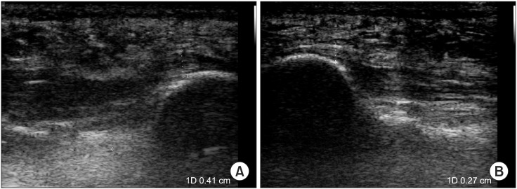
Fig. 2 Ultrasonographic finding of plantar fasciitis. (A) Thickening of the plantar fascia, Sagittal sonogram of 40-year-old man who had a clinical diagnosis of plantar fasciitis. (B) Normal sonographic finding of plantar fascia.
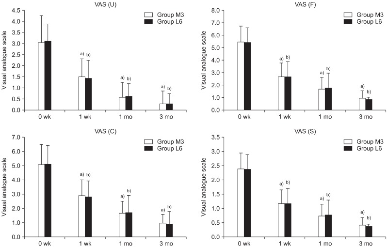
Fig. 3 Temporal changes of pain (mean±standard deviation) at 0 and 1 week and at 1-3 month follow-up after extracorporeal shock wave therapy (ESWT) in the same total energy influx groups (groups M3 and L6). Group M3, 3 sessions of medium-energy (0.16 mJ/mm2) in ESWT group; group L6, 6 session of low-energy (0.08 mJ/mm2) ESWT group; VAS (U), visual analogue scale at usual time; VAS (F), visual analogue scale at first step in the morning; VAS (C), visual analogue scale of compression at calcaneus; VAS (S), visual analogue scale at sleeping. No significant difference was observed between both groups in VAS (U), p=0.717; VAS (F), p=0.778; VAS (C), p=0.829; and VAS (S), p=0.998. a)p<0.001 significantly different vs. 0 week within group M3. b)p<0.001 significantly different vs. 0 week within group L6.
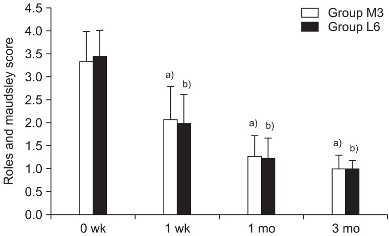
Fig. 4 Temporal changes of Roles and Maudsley score (mean±standard deviation) at 0 and 1 week and at 1-3 months follow-up after extracorporeal shock wave therapy in the same total energy influx groups (groups M3 and L6). a)p<0.001 significantly different vs. 0 week within group M3. b)p<0.001 significantly different vs. 0 week within group L6. No significant difference between both groups (p=0.689) in Roles and Maudsley score.
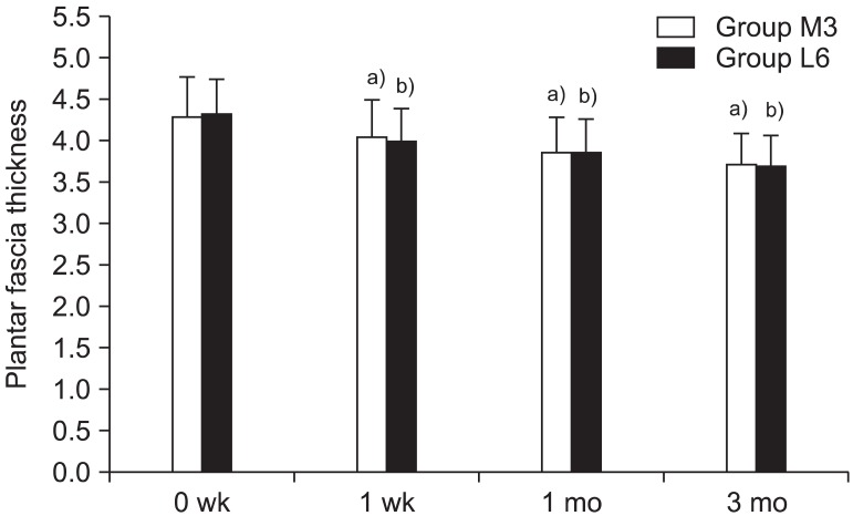
Fig. 5 Temporal changes of plantar fascia thickness (mean±SD) at 0 and 1 week and at 1-3 month follow-up after extracorporeal shock wave therapy in the same total energy influx groups (group M3 and L6). a)p<0.001 significant difference vs. 0 week within group M3. b)p<0.001 significant difference vs. 0 week within group L6. No significant difference occurred between both groups (p=0.859) in plantar fascia thickness.
Cited by 1 articles
-
Therapeutic Effect of Extracorporeal Shock Wave Therapy According to Treatment Session on Gastrocnemius Muscle Spasticity in Children With Spastic Cerebral Palsy: A Pilot Study
Dong-Soon Park, Dong Rak Kwon, Gi-Young Park, Michael Y. Lee
Ann Rehabil Med. 2015;39(6):914-921. doi: 10.5535/arm.2015.39.6.914.
Reference
-
1. Kane D, Greaney T, Shanahan M, Duffy G, Bresnihan B, Gibney R, et al. The role of ultrasonography in the diagnosis and management of idiopathic plantar fasciitis. Rheumatology. 2001; 40:1002–1008. PMID: 11561110.
Article2. Bolgla LA, Malone TR. Plantar fasciitis and the windlass mechanism: a biomechanical link to clinical practice. J Athl Train. 2004; 39:77–82. PMID: 16558682.3. Roxas M. Plantar fasciitis: diagnosis and therapeutic considerations. Altern Med Rev. 2005; 10:83–93. PMID: 15989378.4. Ozdemir H, Yilmaz E, Murat A, Karakurt L, Poyraz AK, Ogur E. Sonographic evaluation of plantar fasciitis and relation to body mass index. Eur J Radiol. 2005; 54:443–447. PMID: 15899349.
Article5. Karabay N, Toros T, Hurel C. Ultrasonographic evaluation in plantar fasciitis. J foot Ankle Surg. 2007; 46:442–446. PMID: 17980840.
Article6. Schepsis AA, Leach RE, Gorzyca J. Plantar fasciitis. Etiology, treatment, surgical results, and review of literature. Clin Orthop Relat Res. 1991; (266):185–196. PMID: 2019049.7. Cole C, Seto C, Gazewood J. Plantar fasciitis: evidence-based review of diagnosis and therapy. Am Fam Physician. 2005; 72:2237–2242. PMID: 16342847.8. Gill LH. Plantar fasciitis: diagnosis and conservative management. J Am Acad Orthop Surg. 1997; 5:109–117. PMID: 10797213.
Article9. Neufeld SK, Cerrato R. Plantar fasciitis: evaluation and treatment. J Am Acad Orthop Surg. 2008; 16:338–346. PMID: 18524985.
Article10. Alvarez RG, Ogden JA, Jaakkola J, Cross GL. Symptom duration of plantar fasciitis and the effectiveness of orthotripsy. Foot Ankle Int. 2003; 24:916–921. PMID: 14733347.
Article11. Ogden JA, Alvarez RG, Marlow M. Shockwave therapy for chronic proximal plantar fasciitis: a meta-analysis. Foot Ankle Int. 2002; 23:301–308. PMID: 11991474.
Article12. Rompe JD, Schoellner C, Nafe B. Evaluation of low-energy extracorporeal shock-wave application for treatment of chronic plantar fasciitis. J Bone Joint Surg Am. 2002; 84:335–341. PMID: 11886900.
Article13. Hammer DS, Rupp S, Kreutz A, Pape D, Kohn D, Seil R. Extracorporeal shockwave therapy (ESWT) in patients with chronic proximal plantar fasciitis. Foot Ankle Int. 2002; 23:309–313. PMID: 11991475.
Article14. Liang HW, Wang TG, Chen WS, Hou SM. Thinner plantar fascia predicts decreased pain after extracorporeal shock wave therapy. Clin Orthop Relat Res. 2007; 460:219–225. PMID: 17353798.
Article15. Hammer DS, Adam F, Kreutz A, Rupp S, Kohn D, Seil R. Ultrasonographic evaluation at 6-month follow-up of plantar fasciitis after extracorporeal shock wave therapy. Arch Orthop Trauma Surg. 2005; 125:6–9. PMID: 14530990.
Article16. Roles NC, Maudsley RH. Radial tunnel syndrome: resistant tennis elbow as a nerve entrapment. J Bone Joint Surg Br. 1972; 54:499–508. PMID: 4340924.17. Pfeffer G, Bacchetti P, Deland J, Lewis A, Anderson R, Davis W, et al. Comparison of custom and prefabricated orthoses in the initial treatment of proximal plantar fasciitis. Foot Ankle Int. 1999; 20:214–221. PMID: 10229276.
Article18. Wolgin M, Cook C, Graham C, Mauldin D. Conservative treatment of plantar heel pain: long-term follow-up. Foot Ankle Int. 1994; 15:97–102. PMID: 7951946.
Article19. Gill LH. Plantar fasciitis: diagnosis and conservative management. J Am Acad Orthop Surg. 1997; 5:109–117. PMID: 10797213.
Article20. Graham CE. Painful heel syndrome: rationale of diagnosis and treatment. Foot Ankle. 1983; 3:261–267. PMID: 6222950.
Article21. Mizel MS, Marymont JV, Trepman E. Treatment of plantar fasciitis with night splint and shoe modifications consisting of a steel shank and anterior rocker bottom. Foot Ankle Int. 1996; 17:732–735. PMID: 8973894.22. Shikoff MD, Figura MA, Postar SE. A retrospective study of 195 patients with heel pain. J Am Podiatr Med Assoc. 1986; 76:71–75. PMID: 3510294.
Article23. Theodore GH, Buch M, Amendola A, Bachmann C, Fleming LL, Zingas C. Extracorporeal shock wave therapy for the treatment of plantar fasciitis. Foot Ankle Int. 2004; 25:290–297. PMID: 15134608.
Article24. Speed CA. Extracorporeal shock-wave therapy in the management of chronic soft-tissue conditions. J Bone Joint Surg Br. 2004; 86:165–171. PMID: 15046427.
Article25. Thiel M. Application of shock waves in medicine. Clin Orthop Relat Res. 2001; (387):18–21. PMID: 11400881.
Article26. Wang CJ, Wang FS, Yang KD, Weng LH, Hsu CC, Huang CS, et al. Shock wave therapy induces neovascularization at the tendon-bone junction: a study in rabbits. J Orthop Res. 2003; 21:984–989. PMID: 14554209.
Article27. Buchbinder R, Ptasznik R, Gordon J, Buchanan J, Prabaharan V, Forbes A. Ultrasound-guided extracorporeal shock wave therapy for plantar fasciitis: a randomized controlled trial. JAMA. 2002; 288:1364–1372. PMID: 12234230.28. Speed CA, Nichols D, Wies J, Humphreys H, Richards C, Burnet S, et al. Extracorporeal shock wave therapy for plantar fasciitis: a double blind randomised controlled trial. J Orthop Res. 2003; 21:937–940. PMID: 12919884.
Article29. Dorotka R, Sabeti M, Jimenez-Boj E, Goll A, Schubert S, Trieb K. Location modalities for focused extracorporeal shock wave application in the treatment of chronic plantar fasciitis. Foot Ankle Int. 2006; 27:943–947. PMID: 17144957.
Article30. Rompe JD, Meurer A, Nafe B, Hofmann A, Gerdesmeyer L. Repetitive low-energy shock wave application without local anesthesia is more efficient than repetitive low-energy shock wave application with local anesthesia in the treatment of chronic plantar fasciitis. J Orthop Res. 2005; 23:931–941. PMID: 16023010.
Article31. Chow IH, Cheing GL. Comparison of different energy densities of extracorporeal shock wave therapy (ESWT) for the management of chronic heel pain. Clin Rehabil. 2007; 21:131–141. PMID: 17264107.32. Rompe JD, Furia J, Weil L, Maffulli N. Shock wave therapy for chronic plantar fasciopathy. Br Med Bull. 2007; 81-82:183–208. PMID: 17456546.
Article33. Gollwitzer H, Diehl P, von Korff A, Rahlfs VW, Gerdesmeyer L. Extracorporeal shock wave therapy for chronic painful heel syndrome: a prospective, double blind, randomized trial assessing the efficacy of a new electromagnetic shock wave device. J Foot Ankle Surg. 2007; 46:348–357. PMID: 17761319.
Article34. Rompe JD, Kirkpatrick CJ, Kullmer K, Schwitalle M, Krischek O. Dose-related effects of shock waves on rabbit tendo Achillis: a sonographic and histological study. J Bone Joint Surg Br. 1998; 80:546–552. PMID: 9619954.35. Takahashi N, Ohtori S, Saisu T, Moriya H, Wada Y. Second application of low-energy shock waves has a cumulative effect on free nerve endings. Clin orthop Relat Res. 2006; 443:315–319. PMID: 16462457.
Article36. Ohtori S, Inoue G, Mannoji C, Saisu T, Takahashi K, Mitsuhashi S, et al. Shock wave application to rat skin induces degeneration and reinnervation of sensory nerve fibers. Neurosci Lett. 2001; 315:57–60. PMID: 11711214.37. Ciampa AR, de Prati AC, Amelio E, Cavalieri E, Persichini T, Colasanti M, et al. Nitric oxide mediates anti-inflammatory action of extracorporeal shock waves. FEBS Lett. 2005; 579:6839–6845. PMID: 16325181.
Article38. Calcagni M, Chen F, Hogger DC, Lindenblatt N, Keel M, Giovanoli P, et al. Microvascular response to shock wave application in striated skin muscle. J Surg Res. 2011; 171:347–354. PMID: 20444475.
Article39. Kudo P, Dainty K, Clarfield M, Coughlin L, Lavoie P, Lebrun C. Randomized, placebo-controlled, double-blind clinical trial evaluating the treatment of plantar fasciitis with an extracorporeal shockwave therapy (ESWT) device: a North American confirmatory study. J Orthop Res. 2006; 24:115–123. PMID: 16435344.
- Full Text Links
-
- Actions
-
Cited
- CITED
-
- Close
- Share
-
- Similar articles
-
- Extracorporeal Shock Wave Therapy for the Treatment of Refractory Plantar Fasciitis
- The Diagnosis and Treatment of Plantar Fasciitis
- The Effect of Extracorporeal Shock Wave Therapy in Plantar Fasciitis
- Suggestions for Effective Extracorporeal Shock Wave Treatment Methods for Lateral Epicondylitis
- Extracorporeal Shock Wave Therapy in Musculoskeletal Disorders
