Ocular Torsion Measured by Fundus Photographs and Optical Coherent Tomography in Normal Koreans
- Affiliations
-
- 1Department of Ophthalmology, Soonchunhyang University Cheonan Hospital, Soonchunhyang University College of Medicine, Cheonan, Korea. ophdrkim@schmc.ac.kr
- KMID: 2217421
- DOI: http://doi.org/10.3341/jkos.2013.54.7.1091
Abstract
- PURPOSE
To investigate the normal range of ocular torsion in Koreans with no ophthalmologic history using fundus photographs and optical coherence tomography and compare the results of the two methods.
METHODS
Fundus photographs and optical coherence tomography were conducted in 400 eyes of 200 people with no ophthalmologic history. ImageJ(R) was used to measure the center of the optic nerve head to foveal angle with fundus photographs. For optical coherence tomography, the fovea-to-disc alignment function in the computer program was used to automatically calculate the angle. Then, the calculated angles measured by the two different methods were compared.
RESULTS
In fundus photographs, the angle of the fovea from the center of the optic nerve head was 6.26 +/- 2.92 degree in the right eye, 6.65 +/- 2.58 degree in the left eye, and the mean value was 6.47 +/- 2.76 degrees. From the automatic calculation in optical coherence tomography, the angle of the fovea from the center of the optic nerve head was 6.12 +/- 3.00 degree in the right eye, 6.83 +/- 2.70 degree in the left eye, and the mean value was 6.52 +/- 2.83 degrees. There was no statistically significant difference between the results of the two different methods. In addition, no statistically significant difference was observed between the right and left eyes, sexes, or ages.
CONCLUSIONS
When comparing the conventional method of measuring ocular torsion with fundus photographs to optical coherent tomography, the fovea-to-disc alignment function of the optical coherent tomography may be useful to automatically calculate the cyclotorsion.
Figure
-

Figure 1. (A) The measurement of disc foveal distance and angle. ‘a’ is horizontal optic disc center-foveal distance. ‘b’ is vertical optic disc center-foveal distance. ‘θ’ is the angle between the optic disc center and the fovea; tan θ = b/a. (B) The measurement of the optic disc center-foveal angle using ImageJ program. ‘a’ is a horizontal line drawn from the optic disc center. ‘b’ is a line drawn from the optic disc center to the fovea. ‘θ’ is the angle between the line a and b.
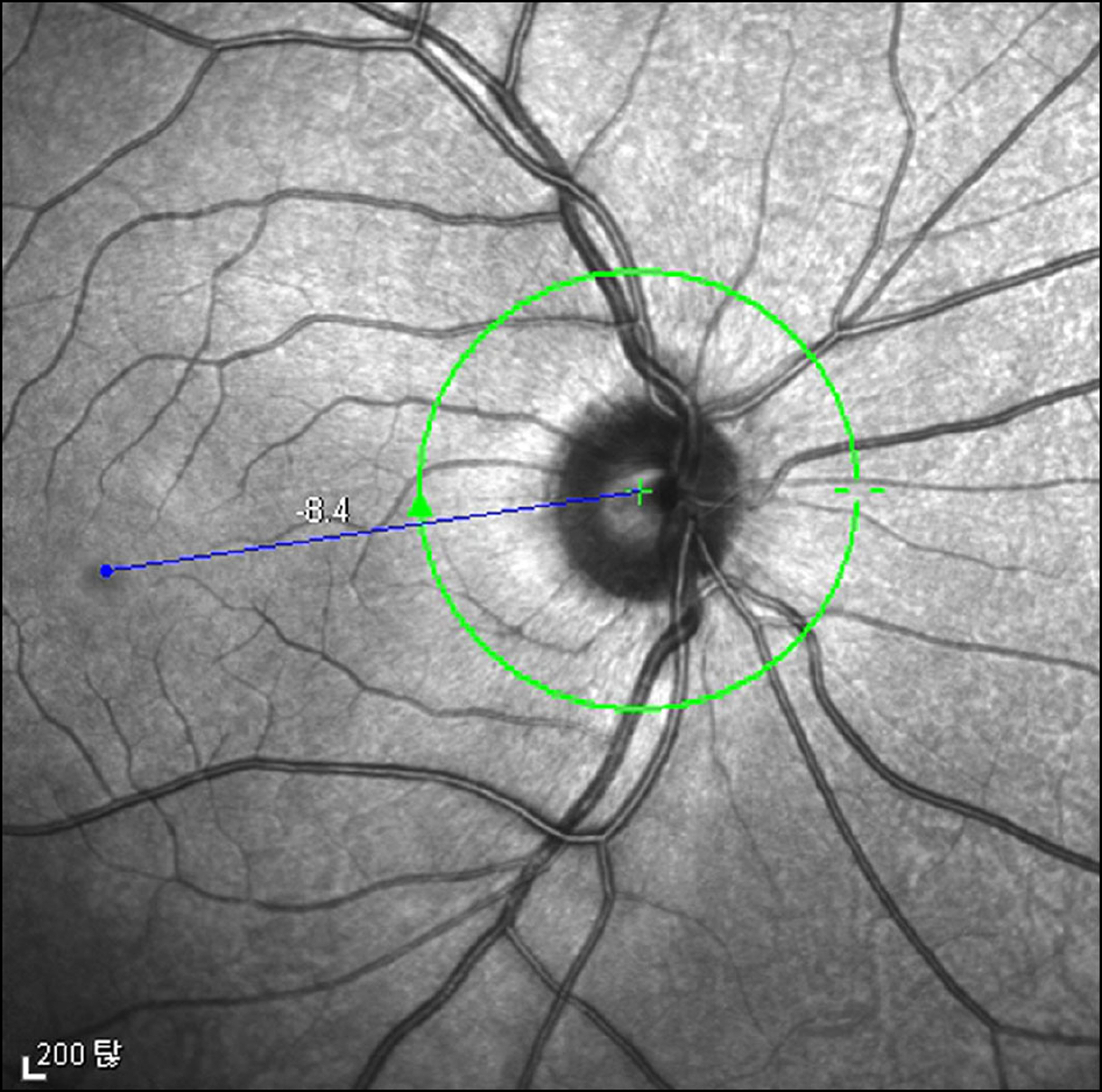
Figure 2. The measurement of the angle between the optic disc center and the fovea using Spectralis optical coherent tomography. Heidelberg eye explorer software provides fo-vea-to-disc alignment function which can accurately measure the angle between the optic disc center and the fovea.
Reference
-
References
1. Kraft SP, O'Reilly C, Quigley PL, et al. Cyclotorsion in unilateral and bilateral superior oblique paresis. J Pediatr Ophthalmol Strabismus. 1993; 30:361–7.
Article2. Bixenman WW, von Noorden GK. Apparent foveal displacement in normal subjects and in cyclotropia. Ophthalmology. 1982; 89:58–62.
Article3. Guyton DL. Clinical assessment of ocular torsion. Am Orthopt J. 1983; 33:7–15.
Article4. von Noorden GK. Clinical observations in cyclodeviations. Ophthalmology. 1979; 86:1451–61.
Article5. von Noorden GK. Clinical and theoretical aspects of cyclotropia. J Pediatr Ophthalmol Strabismus. 1984; 21:126–32.
Article6. Hooten K, Myers E, Worrall R, Stark L. Cyclovergence: the motor response to cyclodisparity. Albrecht Von Graefes Arch Klin Exp Ophthalmol. 1979; 210:65–8.
Article7. Morton GV, Lucchese N, Kushner BJ. The role of funduscopy and fundus photography in strabismus diagnosis. Ophthalmology. 1983; 90:1186–91.
Article8. Jin YH. Location of the foveola in normal subjects. J Korean Ophthalmol Soc. 1985; 26:1019–23.9. Lee DH, Lee SJ, Park SH. Ocular torsion in normal Korean population. J Korean Ophthalmol Soc. 2004; 45:797–802.10. Park SW. The torsional status of normal Koreans. J Korean Ophthalmol Soc. 2004; 45:1906–11.11. Lee HJ, Lim KH. The range of ocular torsion in mass screening. J Korean Ophthalmol Soc. 2005; 46:1684–89.12. Kim EH, Lee SJ, Choi HY. Ocular torsion according to fixation in fundus photograph. J Korean Ophthalmol Soc. 2006; 47:449–54.
- Full Text Links
-
- Actions
-
Cited
- CITED
-
- Close
- Share
-
- Similar articles
-
- Diagnostic Availability of Blind Spot Mapping for Ocular Torsion
- Age and Gender Specific Reference Value of Ocular Torsion by Using Funduscope in Korean
- Clinical Significance of Torsion Measured by Fundus Photography in Children
- Ocular Torsion in Normal Korean Population
- Ocular Torsion according to Fixation in Fundus Photograph
