Macular Ischemia Correlated with Final Visual Outcome in Retinal Vein Occlusion Patients
- Affiliations
-
- 1Department of Ophthalmology, Kosin University College of Medicine, Busan, Korea. hhiatus@gmail.com
- KMID: 2216868
- DOI: http://doi.org/10.3341/jkos.2014.55.10.1493
Abstract
- PURPOSE
To identify the correlation between final visual outcome after at least 6 months of follow-up and the extent of macular ischemia on the first visit.
METHODS
We performed a retrospective clinical analysis of macular ischemia using clinical records, fundus examinations, and fluorescein angiographies in 83 patients (86 eyes) diagnosed with retinal vein occlusion from January 1998 to July 2012 and followed up for over 6 months. We evaluated the extent and the location of macular ischemia, macular edema, initial and final visual acuities and systemic disease based on fluorescein angiography and optical coherence tomography performed within 2 weeks of the first visit. The patients were divided into the following 4 groups based on the extent and location of macular ischemia and edema: superotemporal, superonasal, inferotemporal, and inferonasal.
RESULTS
Retinal vein occlusions (RVOs) consisted of 24 central RVOs (CRVOs) and 62 branch RVOs (BRVOs). Mean initial acuity (log MAR) was 0.35 +/- 0.31 (36 eyes) in the no macular ischemia group, 0.40 +/- 0.21 (11 eyes) in the 1-quadrant macular ischemia group, 0.71 +/- 0.32 (26 eyes) in the 2-quadrant macular ischemia group and 0.73 +/- 0.36 (13 eyes) in the over 3 quadrants macular ischemia group. Mean final acuity (log MAR) was 0.23 +/- 0.23 in the no macular ischemia group, 0.40 +/- 0.30 in the 1-quadrant macular ischemia group, 0.51 +/- 0.32 in the 2-quadrant macular ischemic group and 0.73 +/- 0.31 in the over 3 quadrants macular ischemia group.
CONCLUSIONS
The initial and final visual outcomes were worse when more quadrants were affected by macular ischemia. The extent of macular ischemia was correlated with initial visual acuity and final visual outcome but not with macular edema.
MeSH Terms
Figure
-

Figure 1. (A) Fluorescein angiography shows broken perifoveal capillary network with capillary nonperfusion area which involves 2 quadrants within 1 disc diameter of the center of fovea. (B) Fluorescein angiography (early phase) shows intact perifoveal capillary network without capillary nonperfusion area. (C) Fluorescein angiography shows intact perifoveal capillary network with capillary nonperfusion area at superior m acule.
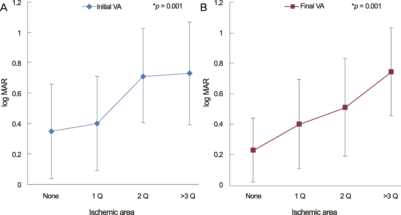
Figure 2. Correlation between the extent of macular ischemia and the visual acuity (VA). (A) As the extent of macular ischemia occupies more area, it shows low initial visual acuity. (B) As the extent of macular ischemia occupies more area, there is a tendency to show low final visual acuity. It means that the larger ischemic area is, the worse visual outcome is to come. Q = quadrant of macular ischemic area. *p-value: statistical significances were tested by one-way ANOVA.
Cited by 1 articles
-
Significance of Early Visual Responses to Bevacizumab for Macular Edema in Branch Retinal Vein Occlusion
Gahyung Ryu, Donghyoun Noh, Junyeop Lee, Min Sagong
J Korean Ophthalmol Soc. 2017;58(8):937-946. doi: 10.3341/jkos.2017.58.8.937.
Reference
-
References
1. Glacet-Bernard A, Coscas G, Chabanel A, et al. Prognostic factors for retinal vein occlusion: prospective study of 175 cases. Ophthalmology. 1996; 103:551–60.2. Kwon OW, Kim JS, Hong YJ, Kim HB. Size of capillary nonperfusion and their prognosis in branch retinal vein occlusion patients. J Korean Ophthalmol Soc. 1993; 34:1162–6.3. Sohn JH, Song SJ. Arteriovenous sheathotomy for persistent macular edema in branch retinal vein occlusion. Korean J Ophthalmol. 2006; 20:210–4.
Article4. Chung JH, Choi GJ, Na KS. The macular circulation state on BRVO according to occlusion site. J Korean Ophthalmol Soc. 2000; 41:1556–62.5. Kang SJ, Chin HS, Moon YS. Visual prognosis of macular edema associated with macular ischemia in branch retinal vein occlusion. J Korean Ophthalmol Soc. 2002; 43:1621–8.6. Lang GE, Freissler K. [Clinical and fluorescein angiography findings in patients with retinal vein occlusion. A unicenter study of 211 patients]. Klin Monbl Augenheilkd. 1992; 201:234–9.7. Park H, Ohn YH, Shin H. Clinical characteristics and classifications of retinal vein occlusion. J Korean Ophthalmol Soc. 1996; 37:1022–31.8. Merin S, Ber I, Ivry M. Retinal ischemia (capillary nonperfusion) in diabetic retinopathy of patients with and without systemic hypertension. Ophthalmologica. 1978; 177:76–81.
Article9. Roseman RL, Olk RJ. Krypton red laser photocoagulation for branch retinal vein occlusion. Ophthalmology. 1987; 94:1120–5.
Article10. Sohn JH, Oh SO, Lee J. The effect of laser treatment on branch retinal vein occlusion. J Korean Ophthalmol Soc. 1993; 34:523–9.11. Kim HC, Kim PS, Kim HK. The macular circulatory change in branch retinal vein occlusion. J Korean Ophthalmol Soc. 1999; 40:1911–7.12. Lee JY, Yoon YH, Kim HK, et al. Baseline characteristics and risk factors of retinal vein occlusion: a study by the Korean RVO study group. J Korean Med Sci. 2013; 28:136–44.
Article13. Clemett RS, Kohner EM, Hamilton AM. The visual prognosis in retinal branch vein occlusion. Trans Ophthalmol Soc UK. 1973; 93:523–35.14. Shilling JS, Jones CA. Retinal branch vein occlusion: a study of argon laser photocoagulation in the treatment of macular oedema. Br J Ophthalmol. 1984; 68:196–8.
Article15. Son MH, Yoon IH. Macular ischemia in temporal branch retinal vein occlusion: fluorescein angiographic classification and visual prognosis. J Korean Ophthalmol Soc. 1996; 37:608–13.16. Finkelstein D. Ischemic macular edema. Recognition and favorable natural history in branch vein occlusion. Arch Ophthalmol. 1992; 110:1427–34.
Article17. Chen SD, Sundaram V, Lochhead J, Patel CK. Intravitreal triamcinolone for the treatment of ischemic macular edema associated with branch retinal vein occlusion. Am J Ophthalmol. 2006; 141:876–83.
Article18. Classification of diabetic retinopathy from fluorescein angiograms. ETDRS report number 11. Early Treatment Diabetic Retinopathy Study Research Group. Ophthalmology. 1991; 98:807–22.19. Shukla D, Kolluru CM, Singh J, et al. Macular ischaemia as a marker for nephropathy in diabetic retinopathy. Indian J Ophthalmol. 2004; 52:205–10.20. Min WK, Hong ST, Park YG, Park KS. The natural course and visual prognosis of branch retinal vein occlusion. J Korean Ophthalmol Soc. 1994; 35:664–72.
- Full Text Links
-
- Actions
-
Cited
- CITED
-
- Close
- Share
-
- Similar articles
-
- Macular Ischemia in Temporal Branch Retinal Vein Occlusion: Fluorescein angiographic classification and Visual prognosis
- Visual Prognosis of Macular Edema Associated with Macular Ischemia in Branch Retinal Vein Occlusion
- Clinical Aspect of Branch Retinal Vein Occlusion
- The Macular Circulation State on BRVO According to Occlusion Site
- Clinical Features According to the Occlusion Site in Patients with Branch Retinal Vein Occlusion
