A Case of Intratarsal Keratinous Cyst of the Meibomian Gland
- Affiliations
-
- 1Department of Ophthalmology, Hallym University Sacred Heart Hospital, Hallym University College of Medicine, Anyang, Korea. tweeti2@hanmail.net
- 2Department of Pathology, Hallym University Sacred Heart Hospital, Hallym University College of Medicine, Anyang, Korea.
- KMID: 2216162
- DOI: http://doi.org/10.3341/jkos.2015.56.1.109
Abstract
- PURPOSE
To report a patient presenting with an intratarsal keratinous cyst of the Meibomian gland in the upper eyelid and a review of the relevant literature.
CASE SUMMARY
A 65-year-old male presented with a right upper eyelid mass which started 5 months prior. The patient reported that the mass recurred several weeks prior even after incision and curettage procedure. The mass was 9 x 5 mm in size and located in the center of the right upper eyelid at the level of lid crease, fixed to the tarsus and a whitish elevated focus was observed at the palpebral conjunctival surface. The mass was excised under local anesthesia and originated from the tarsus. The histopathological examinations revealed an intratarsal keratinous cyst composed of stratified squamous epithelium without keratohyalin granules and filled with keratin. The immunohistochemical studies showed positive staining results for cytokeratin 5/6, epithelial membrane antigen, and carcinoembryonic antigen.
CONCLUSIONS
Intratarsal keratinous cyst of the Meibomian gland should be considered as a differential diagnosis of a recurrent tarsal mass.
MeSH Terms
Figure
-
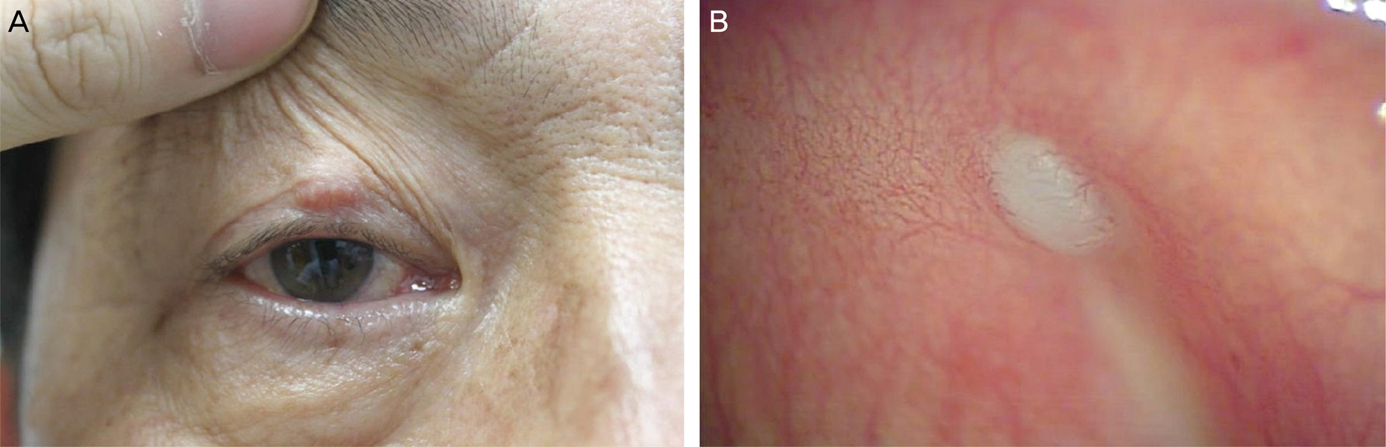
Figure 1. (A) Gross appearance of a mass in the right upper eyelid. The mass was nodular, 9 × 5 mm in size, and fixed to the tarsus, whereas the overlying skin was freely movable. (B) On eversion of the upper eyelid, the tarsal portion of the mass was visible as a slightly elevated and whitish lesion.
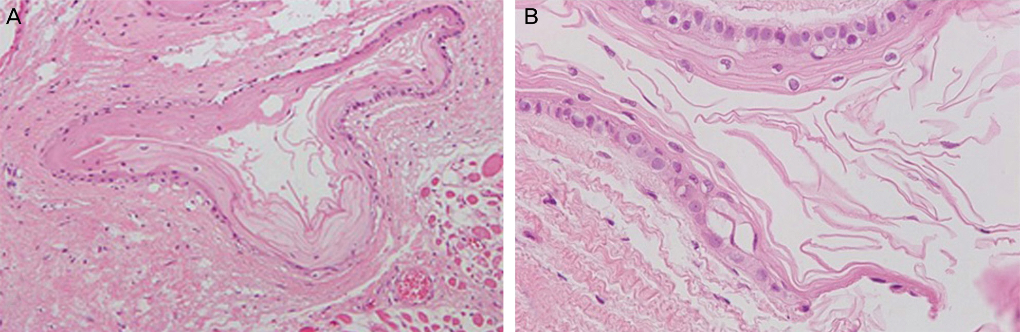
Figure 2. Histopathological findings of the mass. (A) The cyst was lined with stratified squamous epithelium (H&E, magnification ×100). (B) The squamous epithelium of the cystic wall lacks a keratohyalin granular layer. The cystic lumen was filled with keratin strands (H&E, magnification ×400).

Figure 3. (A) Theimmunohistochemical result showed strong positivity for cytokeratin 5/6 (CK-5/6) in the basal and suprabasal layers of the squamous epithelial lining and the luminal keratin strands. (B) Immunoreaction of CK-7 was negative in the cystic wall and keratin. (C) Epithelial membrane antigen is positive in the apical layer of cystic wall and keratin strands, whereas (D) carci-noembryonic antigen is more intensely positive in these regions (magnification ×200).
Reference
-
References
1. Kronish JW, Sneed SR, Tse DT. Epidermal cysts of the eyelid. Arch Ophthalmol. 1988; 106:270–270.
Article2. Jakobiec FA. Sebaceous adenoma of the eyelid and visceral malignancy. Am J Ophthalmol. 1974; 78:952–60.
Article3. Jakobiec FA, Mehta M, Iwamoto M. . Intratarsal keratinous cysts of the Meibomian gland: distinctive clinicopathologic and immunohistochemical features in 6 cases. Am J Ophthalmol. 2010; 149:82–94.
Article4. Lucarelli MJ, Ahn HB, Kulkarni AD, Kahana A. Intratarsal epidermal inclusion cyst. Ophthal Plast Reconstr Surg. 2008; 24:357–9.
Article5. Vagefi MR, Lin CC, McCann JD, Anderson RL. Epidermoid cyst of the upper eyelid tarsal plate. Ophthal Plast Reconstr Surg. 2008; 24:323–4.
Article6. Patel VS, Meyer DR, Carlson JA. Intratarsal keratinous cysts of the meibomian gland (a sebaceous duct cyst): report of 2 cases. Am J Dermatopathol. 2011; 33:624–7.
Article7. Kim HJ, Wojno TH, Grossniklaus HE. Multiple intratarsal keratinous cysts of the eyelid. Ophthal Plast Reconstr Surg. 2012; 28:e116.
Article8. Majumdar M, Khandelwal R, Wilkinson A. Giant epidermal cyst of the tarsal plate. Indian J Ophthalmol. 2012; 60:211–3.
Article9. Gologorsky D, Jakobiec FA, Freitag SK. Transconjunctival elimination of keratin from an intratarsal meibomian cyst. Ophthal Plast Reconstr Surg. 2013; 29:e88–91.
Article10. Rajaii F, Ghafourian A, Eberhart CG. Intratarsal keratinous cyst - an emerging entity. Case Rep Ophthalmol. 2013; 4:160–4.
Article11. Zhang ZD, Li X, Li M. . Clinicopathological features and surgical treatment of intratarsal keratinous cysts. Am J Dermatopathol. 2013; 35:78–82.
Article12. Jenkins JR, Morgan MB. Dermal cysts: a dermatopathological perspective and histological reappraisal. J Cutan Pathol. 2007; 34:815–29.
Article13. Dutton JJ, Fowler AM, Proia AD. Dermoid cyst of conjunctival origin. Ophthal Plast Reconstr Surg. 2006; 22:137–9.
Article14. Jakobiec FA, Bonanno PA, Sigelman J. Conjunctival adnexal cysts and dermoids. Arch Ophthalmol. 1978; 96:1404–9.
Article
