Central Retinal Vein Occlusion Occurrence in an Eales Disease Patient
- Affiliations
-
- 1Department of Ophthalmology, Chungnam National University School of Medicine, Daejeon, Korea. kimjy@cnu.ac.kr
- KMID: 2215965
- DOI: http://doi.org/10.3341/jkos.2012.53.8.1181
Abstract
- PURPOSE
To report the occurrence of central vein occlusion in an Eales disease patient.
CASE SUMMARY
A 23-year-old man presented with decreased left eye visual acuity and was diagnosed with bilateral Eales disease after ophthalmic evaluations. The patient received laser photocoagulation and visual acuity in his left eye improved 1 month after treatment. He was followed up regularly for 3 years and had no specific eye problems. Subsequently, the patient visited our clinic because of visual disturbance in his right eye. The patient's visual acuity was 0.6 in his right eye, and 1.0 in his left eye. On right eye fundus examination, there were multiple flame shape hemorrhages and retinal vascular tortuosity was observed. Arteriovenous transit time was extended on fluorescein angiography. Therefore, the patient was diagnosed with central retinal vein occlusion and underwent an internal medical examination to reveal a possible systemic cause of the central retinal vein occlusion; however, there were no systemic problems. Macular edema was observed on optical coherence tomography and the patient received an intravitreal bevacizumab injection. Six months after treatment, the right eye visual acuity and macular edema improved.
CONCLUSIONS
Reports of branched retinal vein occlusion on the peripheral retina are common in Eales disease patients. However, the authors experienced and report a case of central retinal vein occlusion occurring in Eales disease.
MeSH Terms
-
Antibodies, Monoclonal, Humanized
Eye
Fluorescein Angiography
Hemorrhage
Humans
Light Coagulation
Macular Edema
Neovascularization, Pathologic
Retina
Retinal Vasculitis
Retinal Vein
Retinal Vein Occlusion
Retinaldehyde
Tomography, Optical Coherence
Veins
Visual Acuity
Young Adult
Bevacizumab
Antibodies, Monoclonal, Humanized
Neovascularization, Pathologic
Retinal Vasculitis
Retinaldehyde
Figure
-
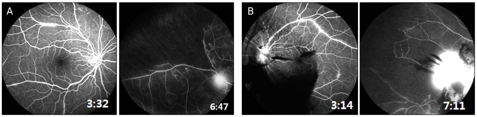
Figure 1 Fluorescein angiography (FA) at initial visit. (A) FA of right eye shows vascular sheathing. New vessel and non-perfusion area are seen on the superotemporal area of retina. (B) Left eye FA shows perivascular leakage and blocked fluorescence due to vitreous hemorrhage. New vessel and non-perfusion area are seen on the superonasal area of the retina.
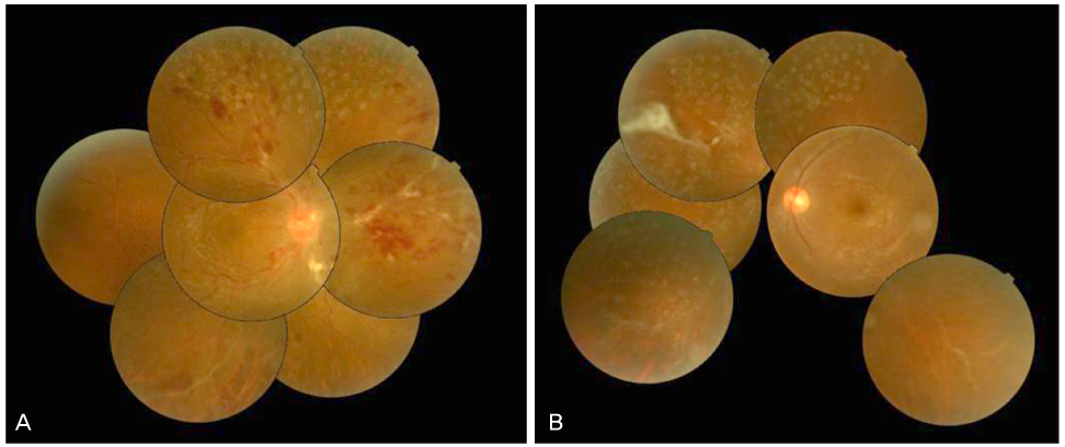
Figure 2 Fundus photography at visit for right eye visual disturbance. (A) The photo of right eyes shows the vessel tortuosity, flame shape hemorrhages, and disc edema. On the periphery, vascular sheaths, retinal hemorrhages, and laser scars are shown. (B) Left fundus photograph shows vascular sheaths, laser scars, and fibrous membrane on the superonasal area.

Figure 3 On Fluorescein angiography of right eye, arteriovenous transit time was delayed and there were diffuse vascular leakage and disc leakage suggesting CRVO (A), peripheral nonperfusion areas and vascular staining which can be shown in Eales disease (B, C).
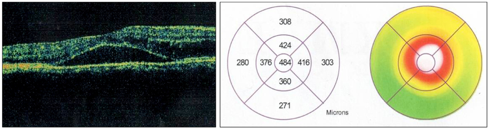
Figure 4 On optical coherence tomography (OCT) of right eye, there is macular edema with subretinal fluid.
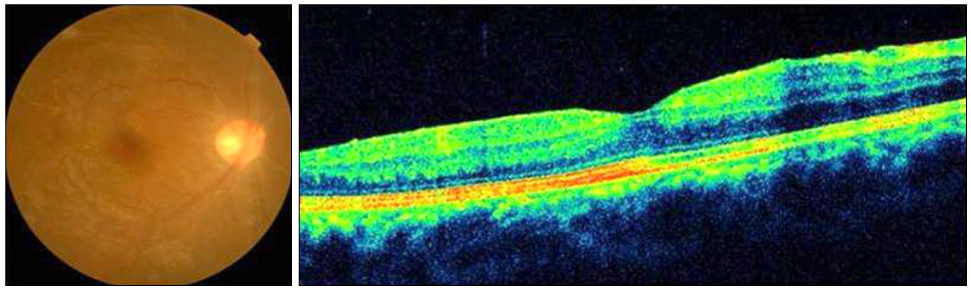
Figure 5 Fundus photography and optical coherence tomography (OCT) of right eye at 6 months after first treatment. There was an improvement of vascular tortuosity, retinal hemorrhage, and macular edema.
Reference
-
1. Biswas J, Sharma T, Gopal L, et al. Eales disease--an update. Surv Ophthalmol. 2002. 47:197–214.2. Moyenin P, Grange JD. [Eales' syndrome. Clinical aspects, therapeutic indications and course of 29 cases]. J Fr Ophtalmol. 1987. 10:123–128.3. Atmaca LS, Idil A, Gündüz K. Visualization of retinal vasculitis in Eales' disease. Ocul Immunol Inflamm. 1993. 1:41–48.4. Das T, Biswas J, Kumar A, et al. Eales' disease. Indian J Ophthalmol. 1994. 42:3–18.5. Renie WA, Murphy RP, Anderson KC, et al. The evaluation of patients with Eales' disease. Retina. 1983. 3:243–248.6. Atmaca LS, Batioglu F, Atmaca Sonmez P. A long-term follow-up of Eales' disease. Ocul Immunol Inflamm. 2002. 10:213–221.7. Gilbert TW. Periphlebitis and endovasculitis of retinal vessels. Klin Monatsbl Augenheilkd. 1935. 94:335–349.8. Gieser SC, Murphy RP. Ryan SJ, editor. Eales disease. Retina. 1989. Vol. 2. St. Louis: Mosby;535–539.9. Ahn JB, Kim YY, Huh K. Eales disease accompanied with branch retinal vein occlusion. J Korean Ophthalmol Soc. 1995. 36:658–663.
- Full Text Links
-
- Actions
-
Cited
- CITED
-
- Close
- Share
-
- Similar articles
-
- Eales Disease Accompanied with Branch Retinal Vein Occlusion
- Clinical Study on The Vitreous Hemorrhage
- Clinical Analysis of the Hemispheric Retinal Vein Occlusion
- A Case of Cilioretinal Artery Occlusion Associated with Central Retinal Vein Occlusion
- Clinical Characteristics and Classifications of Retinal Vein Occlusion
