A New Simple Technique for Removal of Subconjunctival Cyst under the Slit Lamp Microscope
- Affiliations
-
- 1Department of Ophthalmology, Yonsei University College of Medicine, Seoul, Korea.
- 2Siloam Eye Hospital, Seoul, Korea. nairwiny@naver.com
- KMID: 2215161
- DOI: http://doi.org/10.3341/jkos.2011.52.12.1531
Abstract
- PURPOSE
We present a new simple technique to remove subconjunctival cyst under the slit lamp microscope.
CASE SUMMARY
A cotton swab was used to verify whether or not the cyst was freely movable under the conjunctiva. After topical anesthesia, we incised the conjunctiva near the cyst using a 30-gauge needle and extracted the cyst through the wound using forceps under the slit lamp microscope. Four cases of subconjunctival cyst were successfully removed with our new technique. During the average five month (2-10 month) follow-up period, there was no recurrence or procedure-related complications.
CONCLUSIONS
Some subconjunctival cysts such as an epithelial inclusion cyst which is freely movable without attachment to surrounding tissues can be easily removed with a 30-gauge needle and forceps under the slit lamp microscope. This could be considered as the primary procedure instead of simple aspiration.
Figure
-

Figure 1. Case 2 patient. (A) Anterior segment photograph before cyst removal. About 5 × 3 mm sized subconjunctival cyst was noted at nasal region in the left eye. (B) Conjuctival incision was made at the upper margin of the cyst with 30 gauge needle. During the incision, care was taken not to injure the cyst capsule. (C) The cyst was extracted through the wound with forceps. The cyst was removed without damaging cyst capsule. (D) Immediate feature of the conjunctiva after cyst removal. Only small crack was remained.
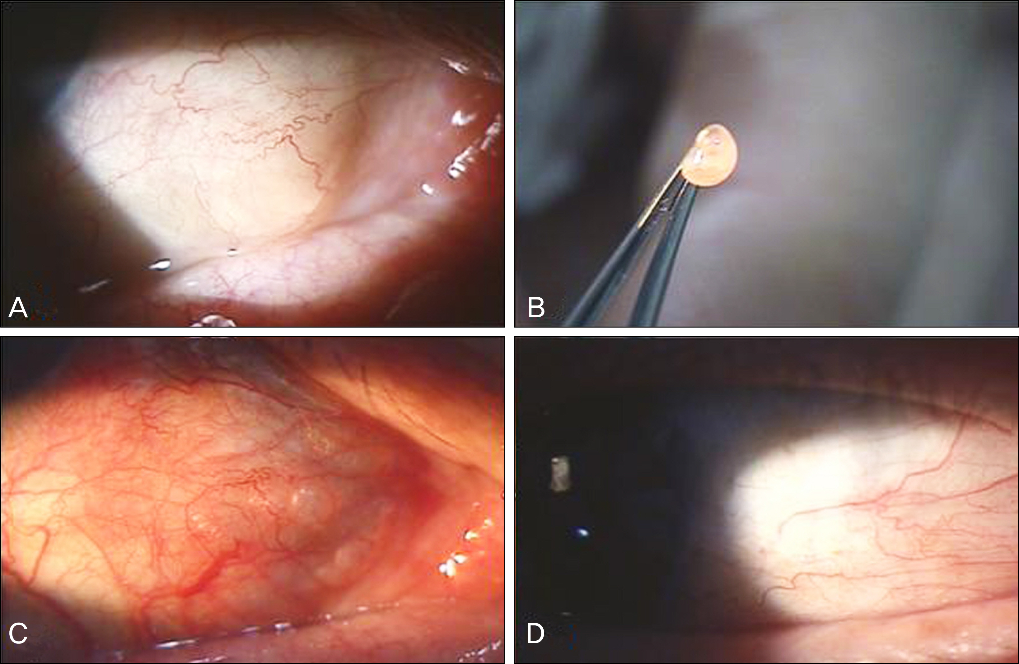
Figure 2. Case 1 patient. (A) About 7 × 7 mm sized round subconjunctival cyst was located at the inferonasal conjunctiva in the right eye. (B) The cyst was removed successfully without the cyst rupture. (C) Immediate feature of the conjunctiva after cyst removal. (D) After 2 month later, there was no recurrence of the cyst.
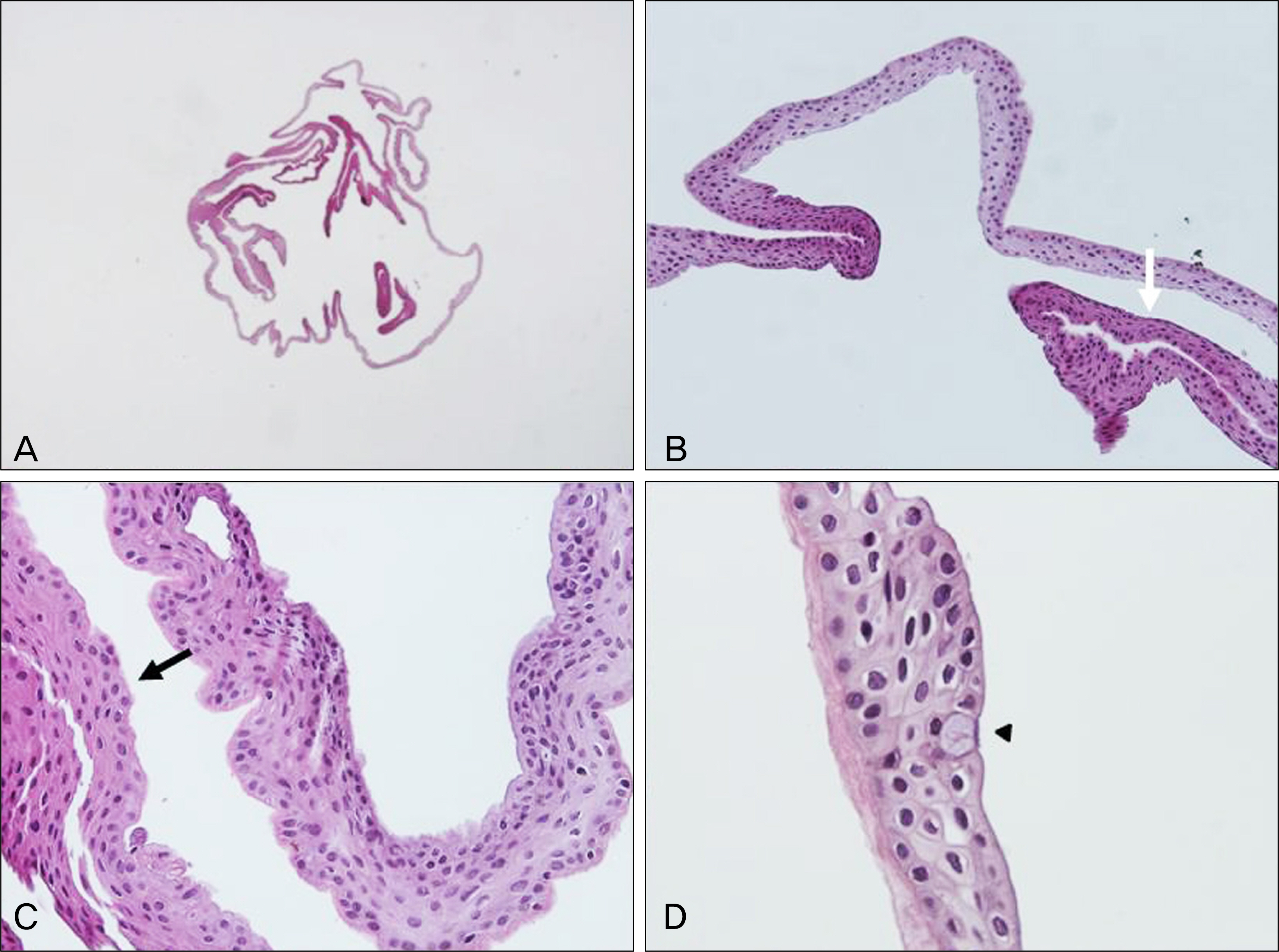
Figure 3. Pathology of the cyst (Case 1 patient). (A) The section revealed that entire epithelial cyst was completely removed without rupture of the capsule (H&E, ×12). (B, C) The wall is consisted of nonkeratizing stratified squamous (white arrow) and cuboidal epithelium (black arrow) (H&E, ×200). (D) Higher power shows the goblet cell (arrow head) in the epithelial lining (H&E, ×400).

Figure 4. Case 3 patient. (A) Extracted subconjunctival cyst: When the conjunctiva was incised, the capsule of cyst was damaged and ruptured. But, we tried to remove subconjunctival tissue through the wound using forceps, and successfully removed underlying tissue. (B) Immediate feature of the conjunctiva after cyst removal. (C) Conjuctival injection was noted after 1 week later after cyst removal. (D) There was no recurrence after 10 month later of the cyst removal.
Cited by 1 articles
-
A Case of Conjunctival Inclusion Cyst Managed with Marsupialization
Hyo Sung Yoon, Min Joung Lee
J Korean Ophthalmol Soc. 2014;55(2):289-292. doi: 10.3341/jkos.2014.55.2.289.
Reference
-
References
1. Shields JA, Shields CL. Tumors of the conjunctiva and cornea. Smolin G, Thoft RA, editors. The Cornea. Scientific Foundations and Clinical Practice. 3rd ed.Boston: Little, Brown;1994. p. 586–95.
Article2. Aponte EP, Schoenfield L, Stern RM, Singh AD. A ductal cyst of lacrimal origin. Advanced Ocular Care. 2010. november/de-cember:29-30.3. Sameshima SS, Beyer-Machule CK. Acquired ptosis associated with a conjunctival cyst. Ophthal Plast Reconstr Surg. 1988; 4:159–62.
Article4. Srinivasan BD, Jakobiec FA, Iwamoto T, DeVoe AG. Epibulbar mucogenic subconjunctival cysts. Arch Ophthalmol. 1978; 96:857–9.
Article5. Brownell RD. Bulbar subconjunctival epithelial cyst. Am J Ophthalmol. 1960; 49:151–3.
Article6. Kotania W, Poniszowska I. Movable cyst in subconjunctival space (in Polish). Klin Oczna. 1969; 39:631–3.7. Bonamour MG, Bonnet M. Subconjunctival mobile cysts. Excision technic. Bull Soc Ophtalmol Fr. 1971; 71:299–300.8. Savar A, Nakra T. Freely mobile subconjunctival cyst. Ophthalmology. 2010; 117:637.e3–4.
Article9. Chan RY, Pong JC, Yuen HK, Lai JS. Use of sodium hyaluronate and indocyanine green for conjunctival cyst excision. Jpn J Ophthalmol. 2009; 53:270–1.
Article10. Kobayashi A, Sugiyama K. Visualization of conjunctival cyst using Healon V and trypan blue. Cornea. 2005; 24:759–60.
Article11. Kobayashi A, Saeki A, Nishimura A, et al. Visualization of conjunctival cyst by indocyanine green. Am J Ophthalmol. 2002; 133:827–8.
Article12. Kobayashi A, Sugiyama K. Successful removal of a large conjunctival cyst using colored 2.3% sodium hyaluronate. Ophthalmic Surg Lasers Imaging. 2007; 38:81–3.
Article
