J Korean Ophthalmol Soc.
2010 Jul;51(7):1023-1027. 10.3341/jkos.2010.51.7.1023.
Surgical Management of Atypical Vogt-Koyanagi-Harada Disease
- Affiliations
-
- 1Department of Ophthalmology, Gyeongsang National University School of Medicine, Jinju, Korea. parkjm@gnu.ac.kr
- 2Institute of Health Science, Gyeongsang National University, Jinju, Korea.
- KMID: 2213796
- DOI: http://doi.org/10.3341/jkos.2010.51.7.1023
Abstract
- PURPOSE
To report a case of surgical treatment of bilateral bullous exudative retinal detachment associated with Vogt-Koyanagi-Harada disease.
CASE SUMMARY
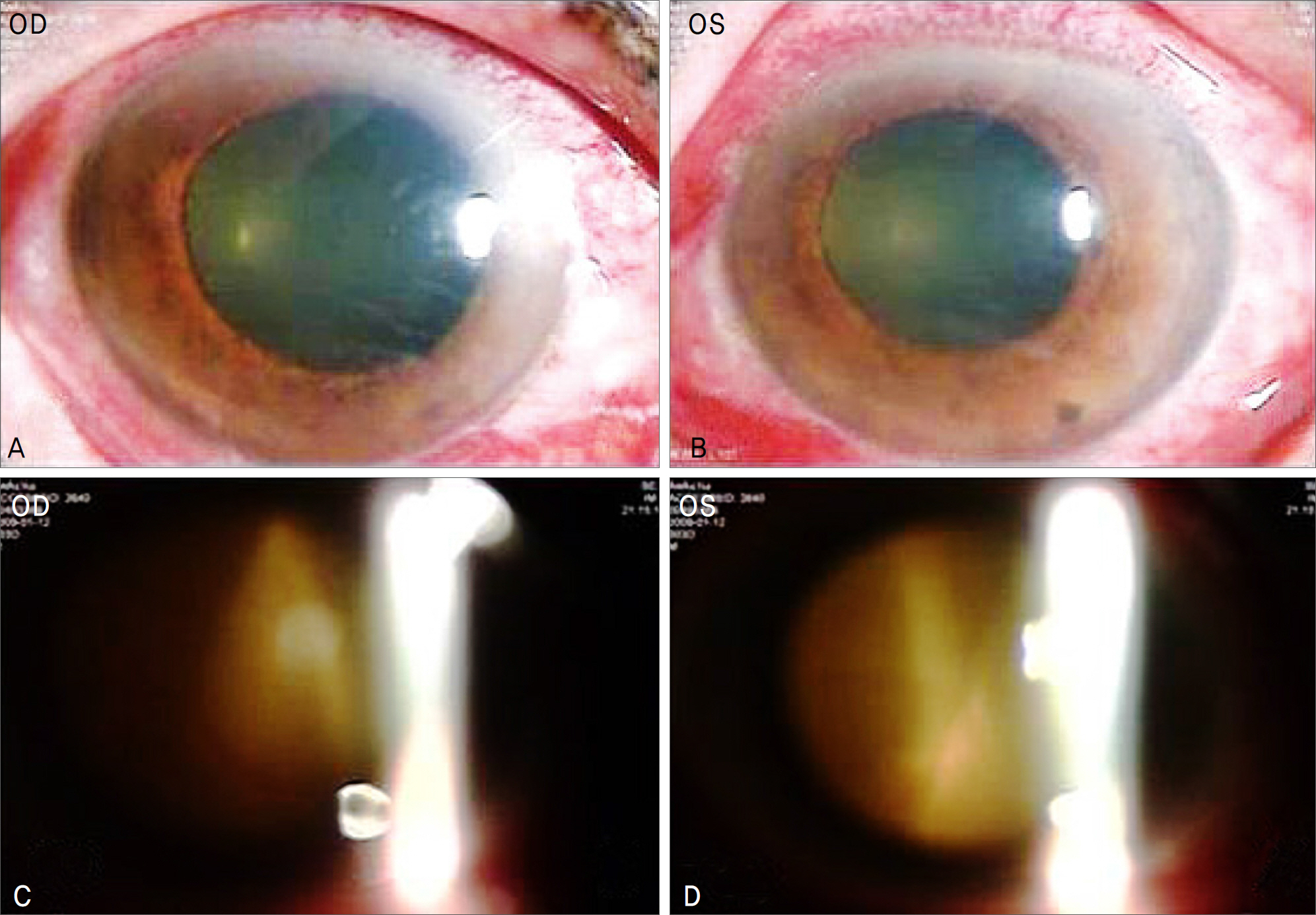
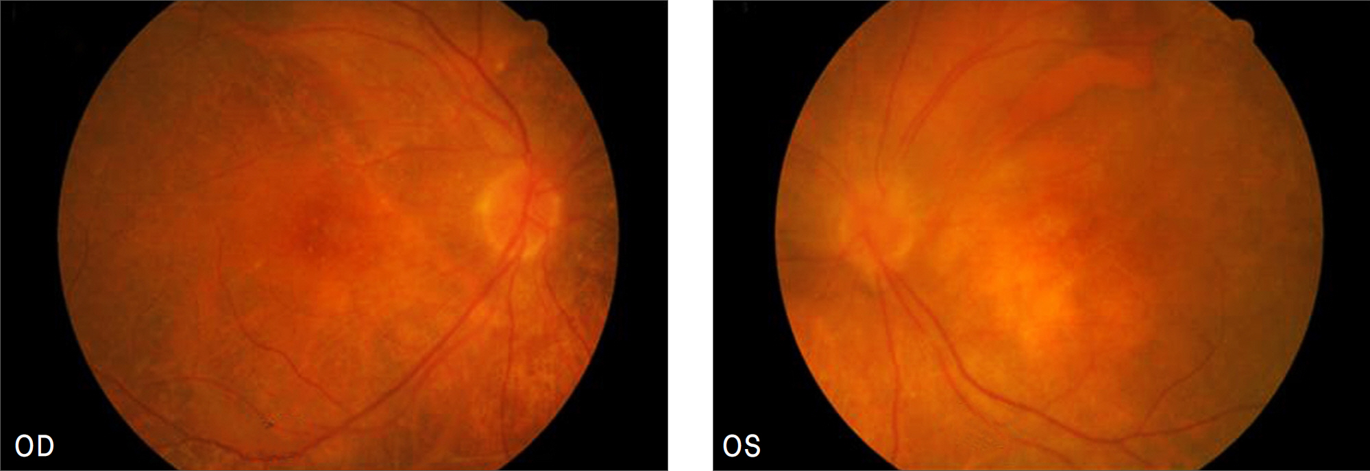
A 64-year-old woman presented with decreased visual acuity, headache, and hearing loss for 2 months. Visual acuity was hand motion in the right eye and light perception in the left eye. Intraocular pressure was 16 mmHg in the right eye and 24 mmHg in the left eye. Slit lamp examimation disclosed corneal edema, conjunctival ciliary injection with chemosis, rubeosis iridis, and posterior synechia in both eyes. Fundus examination demonstrated bilateral bullous exudative retinal detachment. Lumbar puncture revealed pleocytosis and auditory function test showed neurosensory hearing loss. She was diagnosed as having bilateral bullous exudative retinal detachment associated Vogt-Koyanagi-Harada disease. On hospital day 3, intravitreal triamcinolone injection with external subretinal fluid drainage was performed in the right eye and on hospital day 6, intravitreal triamcinolone injection with external subretinal fluid drainage was performed in the left eye. Two months later, best corrected visual acuity was 0.2 in the right eye and 0.04 in the left eye.
CONCLUSIONS
Intravitreal trimacinolone acetonide injection with external subretinal fluid drainage is one of the good treatment for bullous exudative retinal detachment associated with Vogt-Koyanagi-Harada disease.
Keyword
MeSH Terms
Figure
Reference
-
References
1. Kiyomoto C, Imaizumi M, Kimoto K, et al. Vogt–Koyanagi– Harada disease in elderly Japanese patients. Int Ophthalmol. 2007; 27:149–53.2. Kim MJ, Cho NC, Ahn M. Clinical analysis of recurrent Vogt- Koyanagi-Harada syndrome. J Korean Ophthalmol Soc. 2006; 47:227–34.3. Fardeau C, Tran TH, Gharbi B, et al. Retinal fluorescein and in-docyanine green angiography and optical coherence tomography in successive stages of Vogt-Koyanagi-Harada disease. Int Ophthalmol. 2007; 27:163–72.
Article4. Kang JE, Kim HJ, Boo HD, et al. Surgical management of bilateral exudative retinal detachment associated with central serous cho-rio- retinopathy. Korean J Ophthalmol. 2006; 20:131–38.5. Hirose S, Saito W, Yoshida K, et al. Elevated choroidal blood flow ve-locity during systemic corticosteroid therapy in Vogt–Koyanagi– Harada disease. Acta Ophthalmol. 2008; 86:902–7.6. Park JS, Park JM, Lee JE, Oum BS. Vogt-Koyanagi-Harada disease associated with bilateral tonic pupils in a pregnant patient. J Korean Ophthalmol Soc. 2007; 48:1588–92.
Article7. Yamaguchi Y, Otani T, Kishi S. Tomographic features of serous retinal detachment with multilobular dye pooling in acute Vogt- Koyanagi-Harada disease. Am J Ophthalmol. 2007; 144:260–5.8. Gupta V, Gupta A, Gupta P, Sharma A. Spectral-domain cirrus optical coherence tomography of choroidal striations seen in the acute stage of Vogt-Koyanagi-Harada disease. Am J Ophthalmol. 2009; 147:148–53.
Article9. Lim SH, Park SE, Park TK, Ohn YH. Follow-up changes of multi-focal electroretinogram in a patient with Vogt-Koyanaki-Harada syndrome. J Korean Ophthalmol Soc. 2006; 47:494–9.10. Lee SJ, Jin JH, Kim SD. The effect of intravitreal triamcinolone acetonide on intraocular pressure in uveitis. J Korean Ophthalmol Soc. 2007; 48:671–7.11. Seong HK, Lee JM, Park YS, Lee BR. Immediate natural course of IOP after IVTA and the effect of preoperative ocular mass. J Korean Ophthalmol Soc. 2007; 48:808–14.12. Kim WJ, Park YH. The intraocular pressure rise secondary to sub-tenon's injection of triamcinolone after intravitreal injection. J Korean Ophthalmol Soc. 2008; 49:91–7.
Article13. Kim YG, Yu SY, Kwak HW. The effect of intravitreal triamcinolone acetonide injection according to the diabetic macular edema type. J Korean Ophthalmol Soc. 2005; 46:84–9.14. Lee MW, Kyung SE, Chang MH. Prophylactic effect of brimoni-dine 0.15% on IOP elevation after intravitreal triamcinolone acetonide injection. J Korean Ophthalmol Soc. 2008; 49:743–52.
Article15. Kim IT, Park HY, Roh YJ. Bilateral adrenal gland lymphoma masquerading as Vogt-Koyanagi-Harada syndrome. J Korean Ophthalmol Soc. 2008; 49:1198–202.
Article16. Lee SU, Kim SJ, Park YM, et al. Grid laser photocoagulation after intravitreal triamcinolone for macular edema associated with branch retinal vein occlusion. J Korean Ophthalmol Soc. 2009; 50:704–9.
Article17. Kang JE, Kim HJ, Boo HD, et al. Surgical management of bilateral exudative retinal detachment associated with central serous cho-rio- retinopathy. Korean J Ophthalmol. 2006; 20:131–8.


