Relationship Between Visual Acuity and Foveation Window in Infantile Nystagmus by Analyzing Nystagmus Waveforms
- Affiliations
-
- 1Department of Ophthalmology, Soonchunhyang University Bucheon Hospital, Bucheon, Korea.
- 2The Institute of Vision Research, Department of Ophthalmology, Yonsei University College of Medicine, Seoul, Korea. 491209@yuhs.ac
- 3Biosignal Laboratory, Hankyung National University, Suwon, Korea.
- KMID: 2213623
- DOI: http://doi.org/10.3341/jkos.2010.51.6.875
Abstract
- PURPOSE
To report herein on Nystagmus Acuity Estimator Function (NAEF) based on the foveation time, obtained by analyzing waveforms of infantile nystagmus patients and comparing the results with the patients' actual visual acuity.
METHODS
Electro-oculographic data of 27 patients with infantile nystagmus were reviewed. Data of patients only with jerk type nystagmus and reliable visual acuity were analyzed. The foveation time was measured, and NAEF was calculated and compared with the patients' actual best corrected visual acuity.
RESULTS
A correlation analysis of the patients' best corrected visual acuity with NAEF was performed, and the retrieved coefficient was 0.4266. The p-value calculated using the Pearson correlation coefficient was 0.0282, implying that high NAEF correlates positively with visual acuity.
CONCLUSIONS
Estimated visual acuity, calculated based on the waveforms, positively correlates with the patients' actual visual acuity with statistical significance. However, since the foveation time can be measured only in the patients with jerk-type nystagmus waveforms, further study should be performed on the measurement of the foveation time with other waveforms. Furthermore, the present study shows that such analysis is possible with electrooculogram settings in most general hospitals.
Figure
-
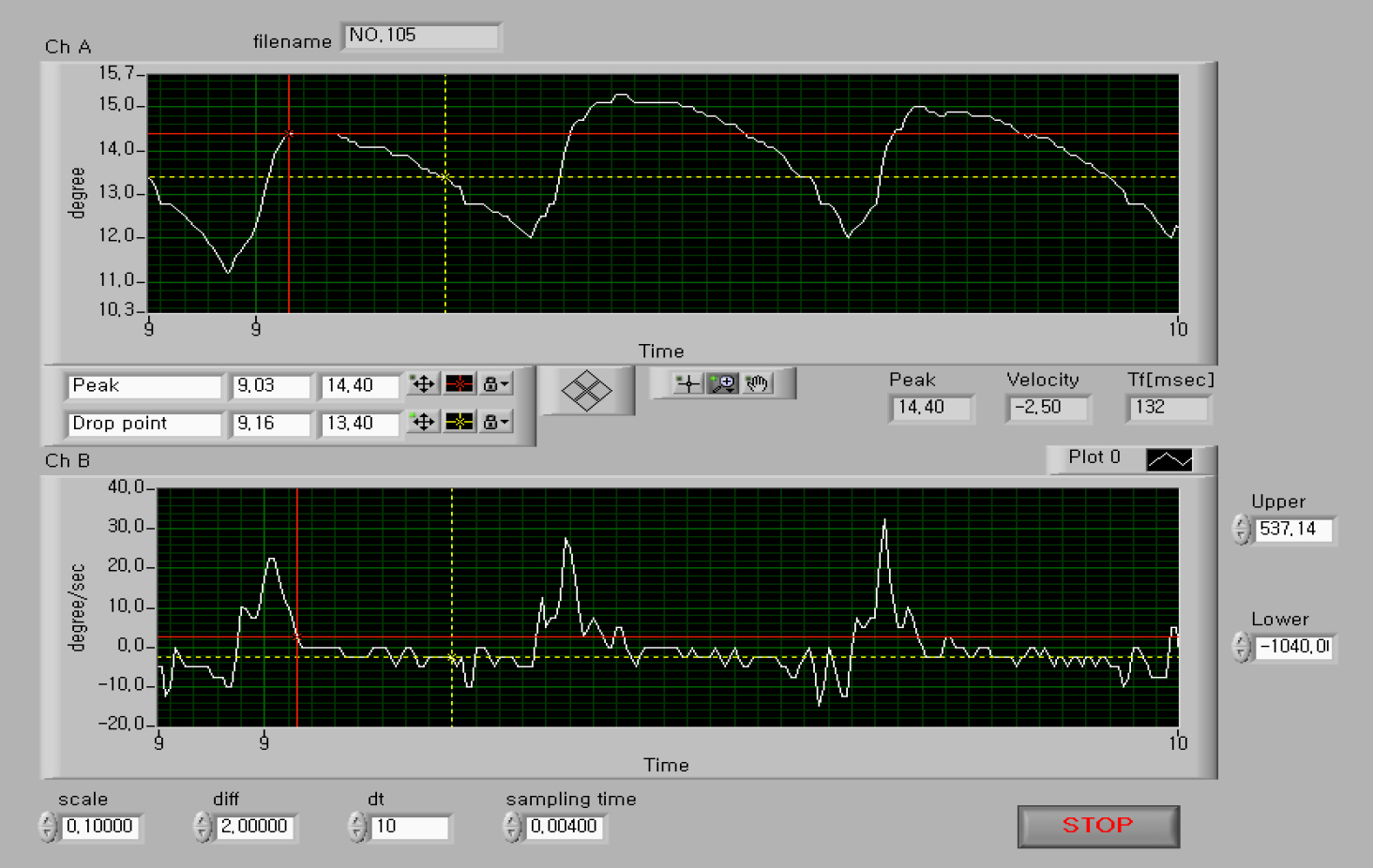
Figure 1. An eye tracing of a 42-year-old male diagnosed with infantile nystagmus shown with our program. Upper graph (Ch A) shows eye positional tracing and lower graph (Ch B) shows eye velocity tracing. Upward deflection denotes rightward direction. First, using cursors (red cross and yellow cross shown on the graph), we identified the interval of ±4°/sec velocity. Second, we check the chosen velocity interval meets the ±0.5° positional criterion. If the interval meets two criteria, we calculate foveation time (Tf) and standard deviation of position (SDp). For de-tails, see the text.
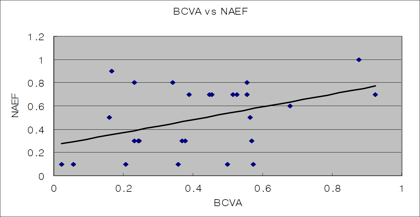
Figure 2. Relationship between NAEF and best corrected visual acuity (BCVA) (correlation coefficient=0.4266. Pearson correlation coefficients: p-value=0.0282) Horizontal axis denotes BCVA (best corrected visual acuity of patients); vertical axis denotes NAEF (nystagmus acuity estimator function of patients).
Cited by 1 articles
-
Laser Refractive Surgery Using an Active Eye-Tracking System in Congenital Nystagmus
Yong Hyun Kim, Sung Yong Kang, Jin Young Choi, Hong Seok Yang, Seung Ah Chung
J Korean Ophthalmol Soc. 2015;56(12):1991-1996. doi: 10.3341/jkos.2015.56.12.1991.
Reference
-
References
1.Dell'Osso LF. Congenital, latent and manifest latent nystagmussimilarities, difference and relation to strabismus. Jap J Ophthalmol. 1985. 29:351–68.2.Dell’Osso LF., Daroff RB. Congenital nystagmus waveforms and foveation strategy. Doc Ophthalmol. 1975. 39:155–82.
Article3.Bedell HE., Loshin DS. Interrelations between measures of visual acuity and parameters of eye movement in congenital nystagmus. Invest Ophthalmol Visual Sci. 1991. 32:416–21.4.Abadi RV., Dickinson CM. Waveform characteristics in congenital nystagmus. Doc Ophthalmol. 1986. 64:153–67.
Article5.Dell'Osso LF., Van Der Steen J., Steinman RM., Collewijn H. Foveation dynamics in congenital nystagmus, I:Fixation. Doc Ophthalmol. 1992. 79:1–23.6.Sheth NV., Dell'Osso LF., Leigh RJ, et al. The effects of afferent stimulation on congenital nystagmus foveation periods. Vision Res. 1995. 35:2371–82.
Article7.Dell’Osso LF., Jacobs JB. An expanded nystagmus acuity function: intra- and intersubject prediction of best-corrected visual acuity. Doc Ophthalmol. 2002. 104:249–76.8.Jacobs JB., Dell’osso LF. Extending the eXpanded Nystagmus Acuity Function for vertical and multiplanar data. Vision Res. 2010. 50:271–8.
Article9.Cesarelli M., Bifulco P., Loffredo L., Bracale M. Relationship between visual acuity and eye position variability during foveations in congenital nystagmus. Doc Ophthalmol. 2000. 101:59–72.10.Keesey UT. Effects of involuntary eye movements on visual acuity. J Opt Soc Am. 1960. 50:769–74.
Article11.Burr DC., Ross J. Contrast sensitivity at high velocities. Vision Res. 1982. 22:479–84.
Article12.Dell'Osso LF. Congenital nystagmus: Basic aspects. Lennerstrand G, Zee DS, Keller EL, editors. Functional basis of ocular motility disorders. 1st ed.New York: Pergamon Press;1982. p. 129–138.13.Bedell HE., White JM., Abplanalp PL. Variability of foveations in congenital nystagmus. Clin Vision Sci. 1989. 4:247–52.14.Ukwade MT., Bedell HE. Variation of congenital nystagmus with viewing distance. Optom Vis Sci. 1992. 69:976–85.
Article15.Curri DC., Bedell FIE and Song S. Visual acuity for optotypes with image motions simulating congenital nystagmus. Clin Vision Sci. 1993. 8:73–84.16.Bifulco P., Cesarelli M., Loffredo L, et al. Eye movement baseline oscil-lation and variability of eye position during foveation in congenital nystagmus. Doc Ophthalmol. 2003. 107:131–6.
