Intraocular Lens Power Calculations Using Dual Scheimpflug Analyzer
- Affiliations
-
- 1Department of Ophthalmology, Kangnam Sacred Heart Hospital, Hallym University College of Medicine, Seoul, Korea. schinn@hanmail.net
- 2Department of Ophthalmology, Seoul National University College of Medicine, Seoul, Korea.
- 3Department of Ophthalmology, Seoul National University Bundang Hospital, Seoul National University College of Medicine, Seongnam, Korea.
- KMID: 2213237
- DOI: http://doi.org/10.3341/jkos.2016.57.3.369
Abstract
- PURPOSE
To investigate the accuracy of intraocular lens power calculations using simulated keratometry (simK) of dual Scheimpflug analyzer and 5 types of formulas in cataract patients.
METHODS
The keratometry (K), axial length (AXL) and anterior chamber depth (ACD) were measured using ultrasound biometry (USB) combined with auto-keratometry (Auto-K), parital coherence interferometry (PCI; IOL master®) and dual Scheimpflug analyzer (DSA; Galilei®) in 39 eyes of 39 patients. Predicted refraction was calculated using Auto-K, mean K of PCI, and simK and total corneal power (TCP) of DSA in the Sanders-Retzlaff-Kraff (SRK-T) formula. The SRK-II, SRK-T, Holladay II, Haigis, and Hoffer-Q formula were used to calculate predicted refraction with the simK of DSA and AXL of USB. Manifest refraction, mean numerical error (MNE) and mean absolute error were evaluated 1, 3 and 6 months after cataract surgery.
RESULTS
TCP of DSA was lower compared with other keratometric values (p < 0.05). The MNE was not different among Auto-K, mean K and simK. The MNE using TCP was larger compared with Auto-K, mean K and simK at 1 month after surgery (p < 0.05). There was a difference in MNE between simK and TCP of DSA at 6 months after surgery (p < 0.05). The MNE of SRK-T formula was the smallest in the intraocular lens (IOL) power calculation using the simK of DSA.
CONCLUSIONS
We suggest using IOL power calculations with simK of DSA and SRK-T formula rather than TCP of DSA in cataract patients with normal corneas.
MeSH Terms
Figure
-
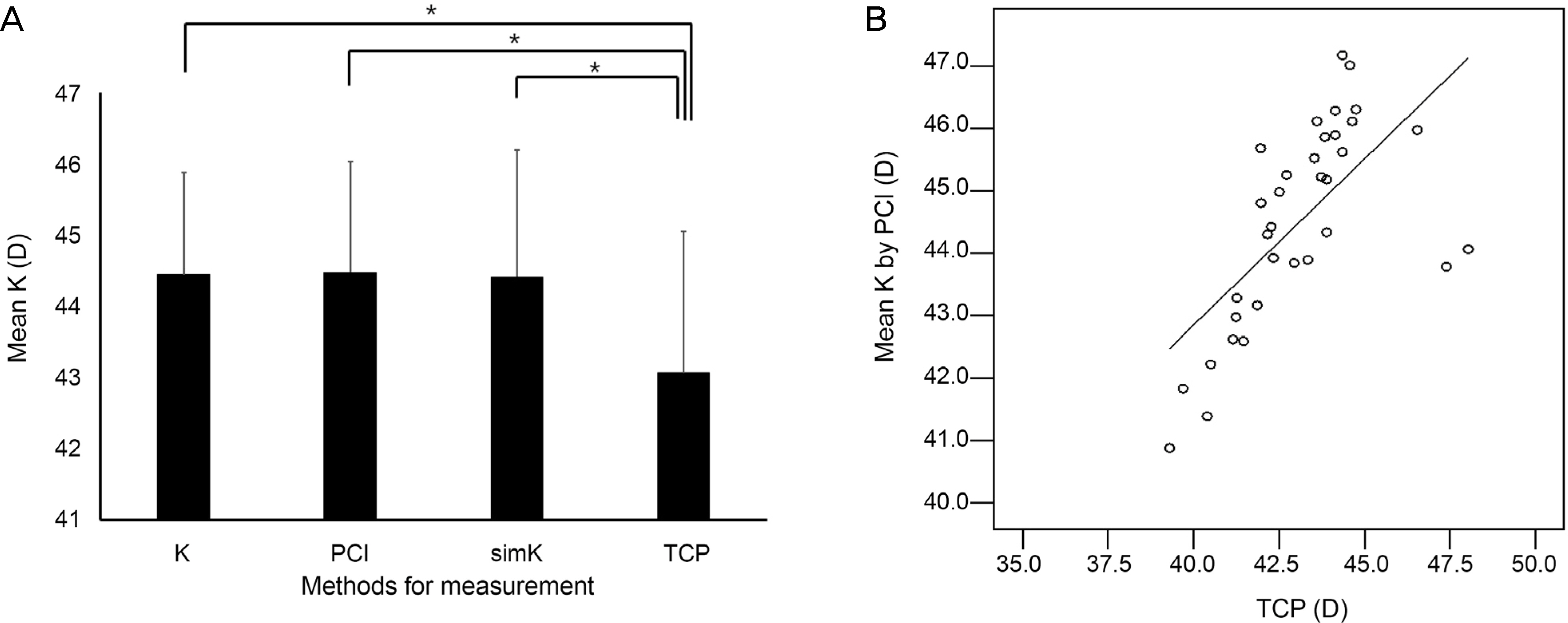
Figure 1. Mean K according to biometry. (A) TCP is low er com pared to auto-keratometry (Auto-K), PCI and simK (p < 0.001 for all, paired t-test). (B) TCP is correlated with PCI (r = 0.653, p < 0.001, Pearson correlation analysis). K = keratometry; PCI = partial coherence interferometry; simK = simulated keratometry; TCP = total corneal power. *Statistically significant.

Figure 2. Correlation between AXL measured by USB and PCI and Bland-Altman plot of axial length between USB and PCI. (A) Axial length measured by USB was well correlated with PCI (r = 0.965, p < 0.001, Pearson correlation analysis). (B) 95% limits of agreement for axial length difference (USB-PCI) is −0.45 to 0.37. PCI = partial coherence interferometry; AXL = axial lengths; USB = ultrasound biometry; SD = standard deviation.
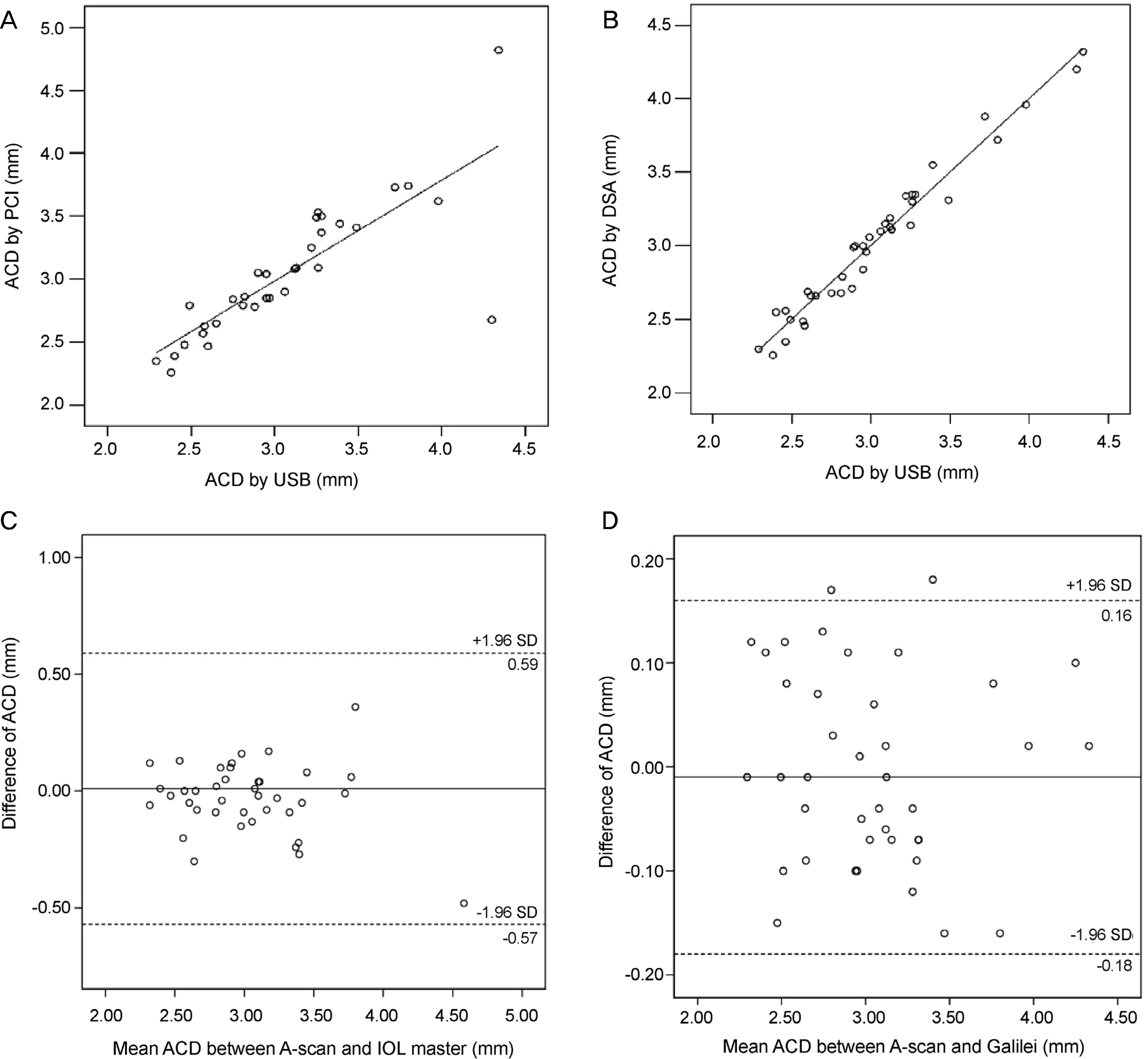
Figure 3. Correlation and Bland-Altman plot of ACD between USB and PCI or DSA. (A) ACD measured by PCI was well correlated with USB (r = 0.811, p < 0.001, Pearson correlation analysis). (B) ACD measured by DSA was well correlated with USB (r = 0.982, p < 0.001, Pearson correlation analysis). (C) 95% limits of agreement for ACD difference (USB-PCI) was −0.57 to 0.59. (D) 95% limits of agreement for ACD difference (USB-DSA) was −0.18 to 0.16. ACD = anterior chamber depth; USB = ultrasound biometry; PCI = partial coherence interferometry; DSA = dual Scheimpflug analyzer; SD = standard deviation.
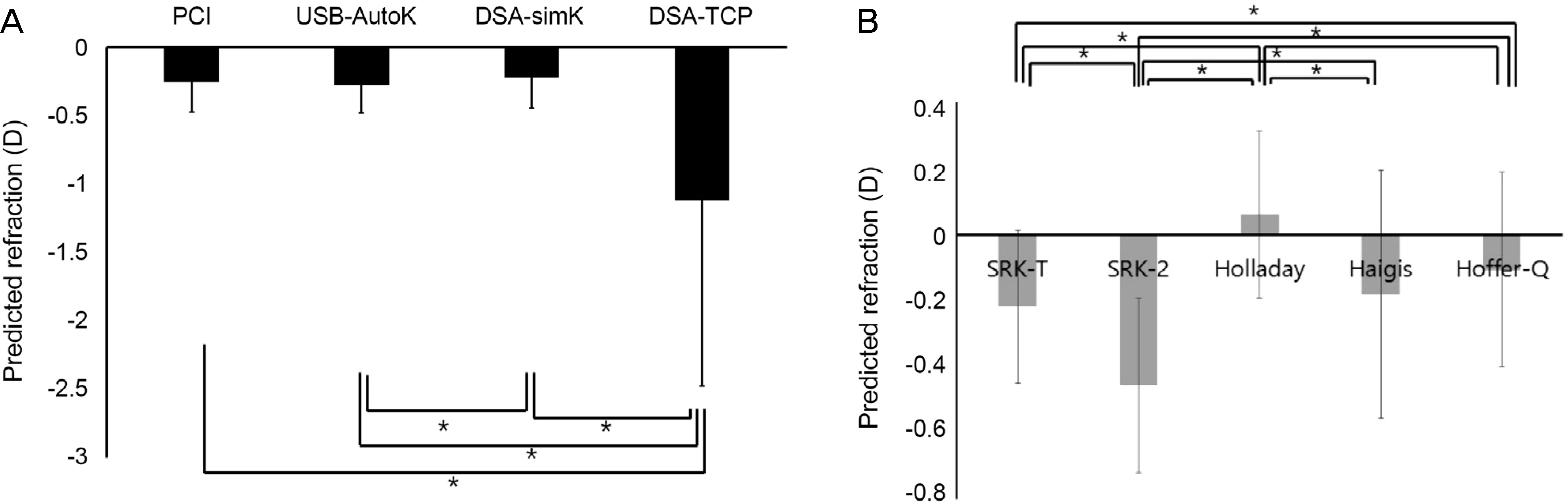
Figure 4. Predicted refraction according to biometry or formula. (A) Predicted refraction is not different between PCI and Auto-K combined with USB or between PCI and DSA. Predicted refraction is different between USB and DSA (p=0.045, paired t-test). Predicted refraction calculated using TCP is higher compared with other methods (p = 0.003 for all, paired t-test). (B) Predicted refraction is different between formulas (p < 0.05, paired t-test). PCI = partial coherence interferometry; Auto-K = auto-keratometry; USB = ultrasound biometry; DSA = dual Scheimpflug analyzer; simK = simulated keratometry; TCP = total corneal power; SRK = Sanders-Retzlaff-Kraff. *Statistically significant.

Figure 5. Change in MRSE overtime. There is no significant change over time (Wilcoxon rank test). MRSE = manifest refraction spherical equivalent.

Figure 6. MNE according to biometry overtime. MNE is not different between PCI, Auto-K combined with USB and DSA 1 month and 3 months after surgery. However, MNE calculated with DSA-simK is lower compared to USB (p = 0.046, Wilcoxon rank test). MNE using TCP is higher compared to other methods 1 month after surgery (p = 0.023, 0.013 and 0.009), and higher compared to DSA-simK 3 months and 6 months after surgery (p = 0.042 and 0.039). MNE = mean numerical error; PCI = partial coherence interferometry; Auto-K = auto-keratometry; USB = ultrasound biometry; DSA = dual Scheimpflug analyzer; simK = simulated keratometry; TCP = total corneal power. *Statistically significant.

Figure 7. MNE according to formula overtime. MNE by SRK-T is lower compared with SRK-2, Holladay or Hoffer-Q formula 1 month after surgery (p = 0.006, < 0.001, and 0.001, respectively, Wilcoxon rank test). MNE by Haigis formula was lower compared with SRK-2, Holladay or Hoffer-Q form ula 1 month after surgery (p < 0.001 for all). SRK-2 and Haigis are lower compared with Holladay formula 1 month after surgery (p < 0.001 for both, respectively). MNE by SRK-T is lower compared with SRK-2, Holladay or Hoffer-Q formula 6 month after surgery (p = 0.014, 0.001, and 0.018, respectively). MNE by Haigis formula is lower compared with SRK-2, Holladay or Hoffer-Q formula 6 month after surgery (p = 0.012, < 0.001 and 0.001, respectively). SRK-2 and Haigis are lower compared with Holladay formula (p < 0.001 and 0.003, respectively). MNE = mean numerical error; SRK-T = Sanders-Retzlaff-Kraff theoretical formula.
Reference
-
References
1. Wang JK, Hu CY, Chang SW. Intraocular lens power calculation using the IOLMaster and various formulas in eyes with long axial length. J Cataract Refract Surg. 2008; 34:262–7.
Article2. Preussner PR, Olsen T, Hoffmann P, Findl O. Intraocular lens calculation accuracy limits in normal eyes. J Cataract Refract Surg. 2008; 34:802–8.3. Drexler W, Findl O, Menapace R, et al. Partial coherence interferometry: a novel approach to biometry in cataract surgery. Am J Ophthalmol. 1998; 126:524–34.
Article4. Olsen T. Sources of error in intraocular lens power calculation. J Cataract Refract Surg. 1992; 18:125–9.
Article5. Lam AK, Chan R, Pang PC. The repeatability and accuracy of axial length and anterior chamber depth measurements from the IOLMaster. Ophthalmic Physiol Opt. 2001; 21:477–83.6. Findl O, Drexler W, Menapace R, et al. High precision biometry of pseudophakic eyes using partial coherence interferometry. J Cataract Refract Surg. 1998; 24:1087–93.
Article7. Haigis W, Lege B, Miller N, Schneider B. Comparison of immersion ultrasound biometry and partial coherence interferometry for intraocular lens calculation according to Haigis. Graefes Arch Clin Exp Ophthalmol. 2000; 238:765–73.
Article8. Holladay JT. Standardizing constants for ultrasonic biometry, keratometry, and intraocular lens power calculations. J Cataract Refract Surg. 1997; 23:1356–70.
Article9. Saad E, Shammas MC, Shammas HJ. Scheimpflug corneal power measurements for intraocular lens power calculation in cataract surgery. Am J Ophthalmol. 2013; 156:460–7.e2.
Article10. Savini G, Barboni P, Carbonelli M, Hoffer KJ. Comparison of methods to measure cornealpower for intraocular lens power calculation using a rotating Scheimpflug camera. J Cataract Refract Surg. 2013; 39:598–604.11. Saiki M, Negishi K, Kato N, et al. Ray tracing software for intraocular lens power calculation after corneal excimer laser surgery. Jpn J Ophthalmol. 2014; 58:276–81.
Article12. Savini G, Barboni P, Carbonelli M, Hoffer KJ. Accuracy of Scheimpflug corneal power measurements for intraocular lens power calculation. J Cataract Refract Surg. 2009; 35:1193–7.
Article13. Minami K, Kataoka Y, Matsunaga J, et al. Ray-tracing intraocular lens power calculation using anterior segment optical coherence tomography measurements. J Cataract Refract Surg. 2012; 38:1758–63.
Article14. Shammas HJ, Hoffer KJ, Shammas MC. Scheimpflug photography keratometry readings for routine intraocular lens power calculation. J Cataract Refract Surg. 2009; 35:330–4.
Article15. Savini G, Barboni P, Carbonelli M, Hoffer KJ. Accuracy of a dual Scheimpflug analyzer and a corneal topography system for intraocular lens power calculation in unoperated eyes. J Cataract Refract Surg. 2011; 37:72–6.
Article16. Shirayama M, Wang L, Koch DD, Weikert MP. Comparison of accuracy of intraocular lens calculations using automated keratometry, a Placido-based corneal topographer, and a combined Placido-based and dual Scheimpflug corneal topographer. Cornea. 2010; 29:1136–8.17. Zaldivar R, Shultz MC, Davidorf JM, Holladay JT. Intraocular lens power calculations in patients with extreme myopia. J Cataract Refract Surg. 2000; 26:668–74.
Article18. Findl O. Biometry and intraocular lens power calculation. Curr Opin Ophthalmol. 2005; 16:61–4.
Article19. Rose LT, Moshegov CN. Comparison of the Zeiss IOLMaster and applanation A-scan ultrasound: biometry for intraocular lens calculation. Clin Experiment Ophthalmol. 2003; 31:121–4.
Article20. Németh J, Fekete O, Pesztenlehrer N. Optical and ultrasound measurement of axial length and anterior chamber depth for intraocular lens power calculation. J Cataract Refract Surg. 2003; 29:85–8.
Article21. Utine CA, Altin F, Cakir H, Perente I. Comparison of anterior chamber depth measurements taken with the Pentacam, Orbscan IIz and IOLMaster in myopic and emmetropic eyes. Acta Ophthalmol. 2009; 87:386–91.
Article22. Reddy AR, Pande MV, Finn P, El-Gogary H. Comparative estimation of anterior chamber depth by ultrasonography, Orbscan II, and IOLMaster. J Cataract Refract Surg. 2004; 30:1268–71.
Article23. Jonna G, Channa P. Updated practical intraocular lens power calculation after refractive surgery. Curr Opin Ophthalmol. 2013; 24:275–80.
Article24. Lee AC, Qazi MA, Pepose JS. Biometry and intraocular lens power calculation. Curr Opin Ophthalmol. 2008; 19:13–7.
Article25. Na JK, Kim MS. The comparison of astigmatic outcomes after cataract surgery of inferior versus superior clear corneal incision. J Korean Ophthalmol Soc. 2014; 55:1470–5.
Article
- Full Text Links
-
- Actions
-
Cited
- CITED
-
- Close
- Share
-
- Similar articles
-
- Astigmatic Correlation between the Automated Refractometry and Dual Scheimpflug Analyzer in Pseudophakic Eyes
- Internal Lens Signal Measured by Dual Scheimpflug Anterior Segment Analyzer
- Comparison of Toric Intraocular Lens Axis Accuracy between Optical Biometry and Dual Scheimpflug Topography
- Intraocular Lens Power Calculation According to the Difference between Anterior and Total Keratometry Using Scheimpflug Imaging
- Comparison of Intraocular Lens Power Calculation Methods Following Myopic Laser Refractive Surgery: New Options Using a Rotating Scheimpflug Camera
