Comparison of the Normal Visual Fields Between the Goldmann and Humphrey Kinetic Perimetries
- Affiliations
-
- 1Department of Ophthalmology, Samsung Medical Center, Sungkyunkwan University School of Medicine, Seoul, Korea.
- 2Department of Ophthalmology, Soonchunhyang University School of Medicine, Seoul, Korea. scheye@hosp.sch.ac.kr
- KMID: 2212369
- DOI: http://doi.org/10.3341/jkos.2009.50.6.904
Abstract
-
PURPOSE: To show Humphrey automated kinetic perimetry can be substituted for Goldmann perimetry, which has been used in the field of disability evaluation field, the differences of normal visual fields between two perimetries were evaluated.
METHODS
Goldmann and Humphrey automated kinetic perimetries were performed simultaneously in 70 eyes of 35 normal healthy Koreans who had no specific ophthalmologic disease at 12 meridians; 0degrees, 30degrees, 60degrees, 90degrees, 120degrees, 150degrees, 180degrees, 210degrees, 240degrees, 270degrees, 300degrees, and 330degrees. The mean values of field in each case were compared. In addition, the corrected values were obtained through the calculation of the difference in the two maximal fields.
RESULTS
The visual fields of Humphrey and Goldmann kinetic perimetries showed a similar oval shape, but the fields of Goldmann were statistically significantly wider than the Humphrey fields. As the values of Humphrey were compared with the original data of Goldmann, all values of the visual field were narrow.
CONCLUSIONS
The visual fields by Humphrey automated kinetic perimetry were smaller than those by Goldmann perimetry. Therefore, if Humphrey kinetic perimetry is used for the evaluation of visual disability, the visual field should be evaluated after the correction.
Figure
-
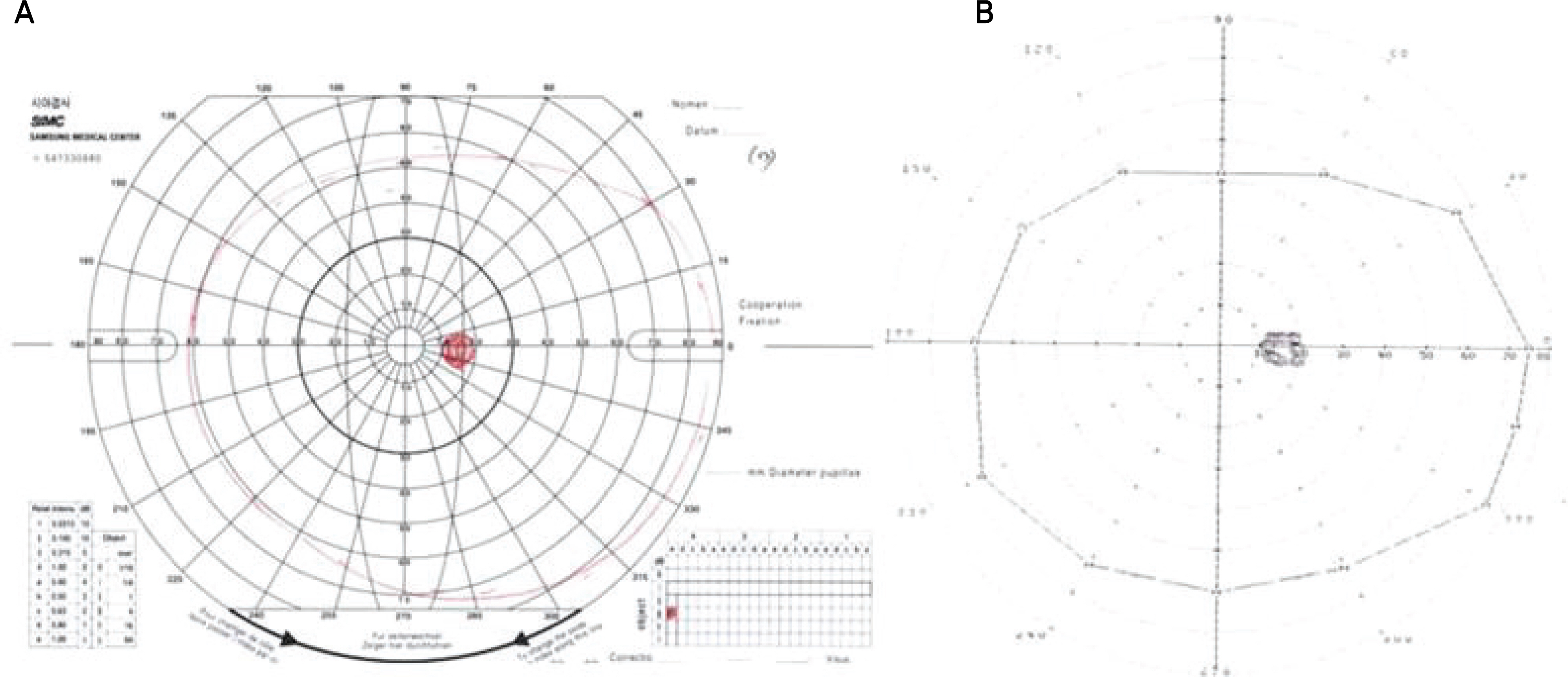
Figure 1. One example of Goldmann perimetry (A) and Humphrey kinetic perimetry (B).

Figure 2. Comparison of Goldmann perimetry and Humphrey kinetic perimetry, right eye (A) and left eye (B).
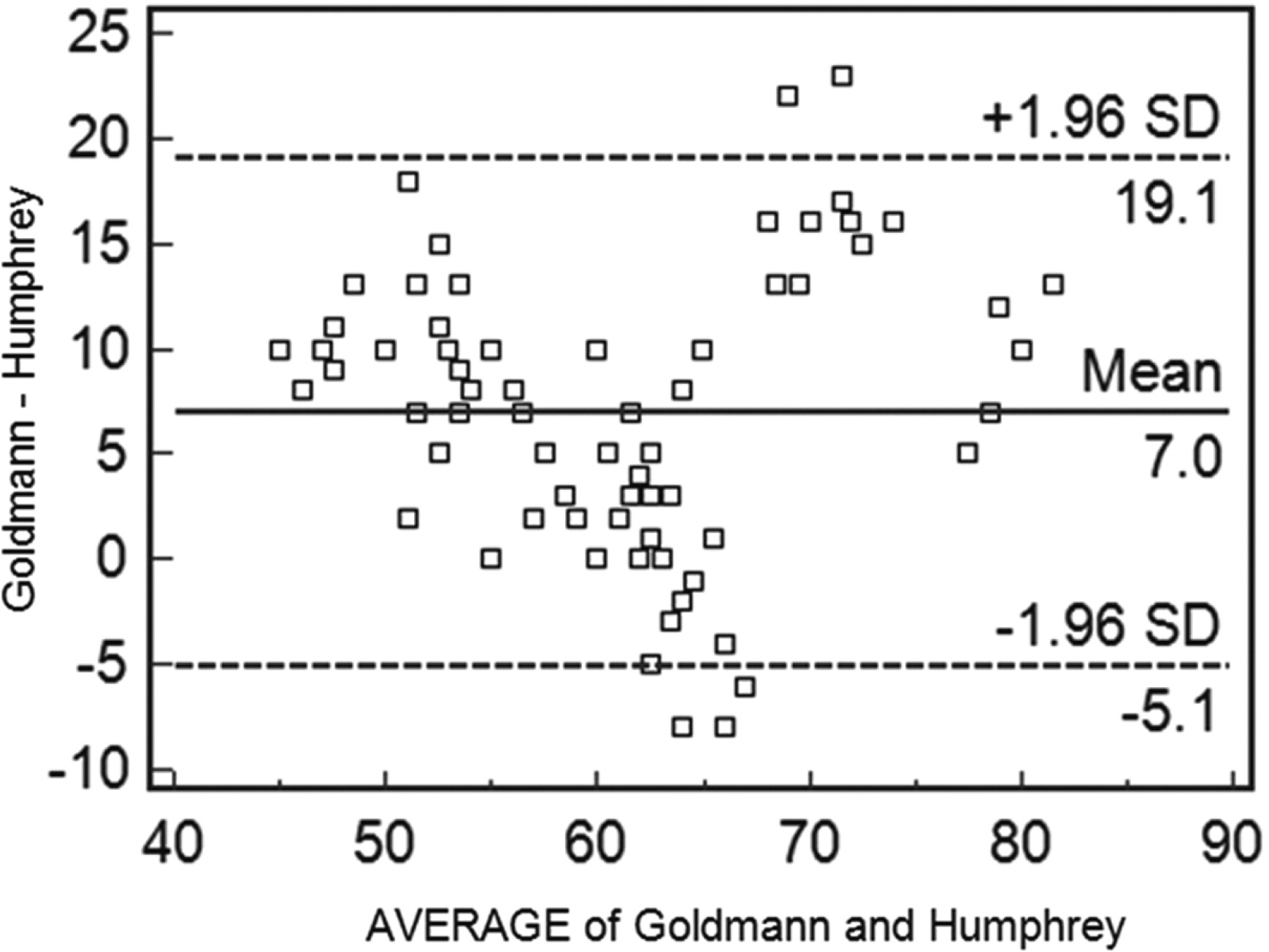
Figure 3. Bland-Altman plot showing the mean values and the error allowance limits of the difference between the values of Goldmann visual field and Humphery visual field.
Cited by 2 articles
-
Comparative Analysis of the Humphrey Static Perimetry and the Goldmann Kinetic Perimetry: Application of the Humphrey Static Perimetry to Visual Disability Evaluation
Jin Hee Shin, Song Hee Park
J Korean Ophthalmol Soc. 2013;54(12):1907-1917. doi: 10.3341/jkos.2013.54.12.1907.A Case of Unusual Pituitary Apoplexy Presented as Aseptic Meningitis
Kang Min Park, Yeon Mee Kim, Si Eun Kim, Kyong Jin Shin, Sam Yeol Ha, Jinse Park, Sung Eun Kim
Korean J Clin Neurophysiol. 2013;15(1):24-26. doi: 10.14253/kjcn.2013.15.1.24.
Reference
-
References
1. Lee SC, Yu SY, Kwak HW. The study of normal visual fields using Goldmann module in OCTOPUS 101 automated perimetry in Koreans. J Korean Ophthalmol Soc. 2004; 45:1123–7.2. Stewart WC, Shields MB. The peripheral visual field in glaucoma: Reevaluation in the age of automated perimetry. Surv Ophthalmol. 1991; 36:59–69.
Article3. Miller KN, Shields MB, Ollie AR. Automated kinetic perimetry with two peripheral isopters in glaucoma. Arch Ophthalmol. 1989; 107:1316–20.
Article4. Miller KN, Shields MB, Ollie AR. Peripheral visual field testing by automated kinetic perimetry in glaucoma. Arch Ophthalmol. 1988; 106:202–6.5. Liu GT, Volpe NJ, Galetta SL. Neuro-Ophthalmology, Diagnosis and Management. Philadelphia: Saunders Co.;2001. p. 46–54.6. Zingirian M, Gandolfo E, Orciuolo M. Automation of the Goldmann perimeter. Doc Ophthalmol Proc Ser. 1983; 42:103–7.
Article7. Kim DK, Choi KS, Park SH. Usefulness of the Binocular Double Vision Field Using Kinetic Automated Perimetry in Diplopia. J Korean Ophthalmol Soc. 2005; 46:1196–203.8. Kim MH, Lee CS. Studies on the normal visual field of Koreans. J Korean Ophthalmol Soc. 1983; 24:367–78.9. Trope GE, Britton R. A comparison of Goldmann and Humphrey automated perimetry in patients with glaucoma. Br J Ophthalmol. 1987; 71:489–93.
Article
- Full Text Links
-
- Actions
-
Cited
- CITED
-
- Close
- Share
-
- Similar articles
-
- Comparative Analysis of the Humphrey Static Perimetry and the Goldmann Kinetic Perimetry: Application of the Humphrey Static Perimetry to Visual Disability Evaluation
- Usefulness of the Binocular Double Vision Field Using Kinetic Automated Perimetry in Diplopia
- The Study of Normal Visual Fields Using Goldmann Module in OCTOPUS 101 Automated Perimetery in Koreans
- Humphrey SITA and Octopus TOP Perimetry on Normal Korean Subjects
- The Influence of Miotics on Visual Field in Glaucoma
