Confocal Microscopic Findings of Transplanted Cornea 10 Years After Penetrating Keratoplasty
- Affiliations
-
- 1Department of Ophthalmology, School of Medicine, Pusan National University Hospital, Busan, Korea. jongsool@pusan.ac.kr
- KMID: 2212324
- DOI: http://doi.org/10.3341/jkos.2009.50.5.779
Abstract
-
PURPOSE:To examine the morphological characteristics of the transplanted cornea in patients who received penetrating keratoplasty at least 10 years previously prior.
CASE SUMMERY: Each layer of the transplanted cornea of patients who received penetrating keratoplasty 10 years previously prior was examined with a confocal microscope (ConfoScan 4.1, Fortune Technology, Italy). Cross-sectioned corneal images of the corneal epithelium, Bowman's layer, stromal layer, Descement's membrane, and endothelium were evaluated and compared with the normal fellow other eye. A total of three eyes from three subjects between the ages of 60 and 70 years were examined. The epithelial cells had large cell borders, and a spindle shaped, and a highly reflective nucleus. The keratocytes were highly reflective and the density of keratocytes was lower than that in the normal cornea. The regenerated nerve fibers were markedly altered, as characterized by increased nerve tortuosity, reduced branching patterns, and shorter nerve lengths. In the endothelial cell layer, a bright nucleus, a reduced ratio of hexagonal cells, and several multinuclear cells were observed.
CONCLUSIONS
Even 10 years after penetrating keratoplasty, the entire transplanted cornea is morphologically different from the normal cornea.
MeSH Terms
Figure
-
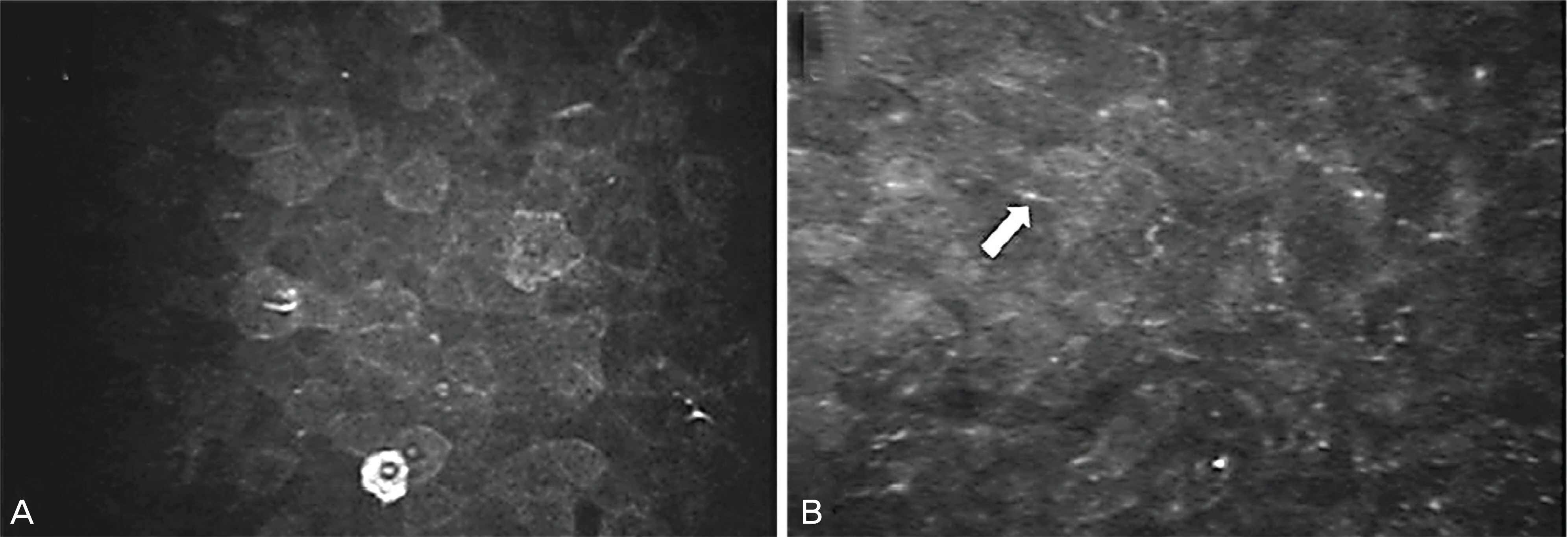
Figure 1. Corneal epithelial layer images of normal human (A) and penetrating keratoplasty patient (B) digitalized directly from confocal microscopy through focusing scan (case 2). (A) normal epithelial cells. (B) abnormally elongated epithelial cells (arrow) were found in the cornea after PKP. The nucleus of epithelial cells was more prominent.
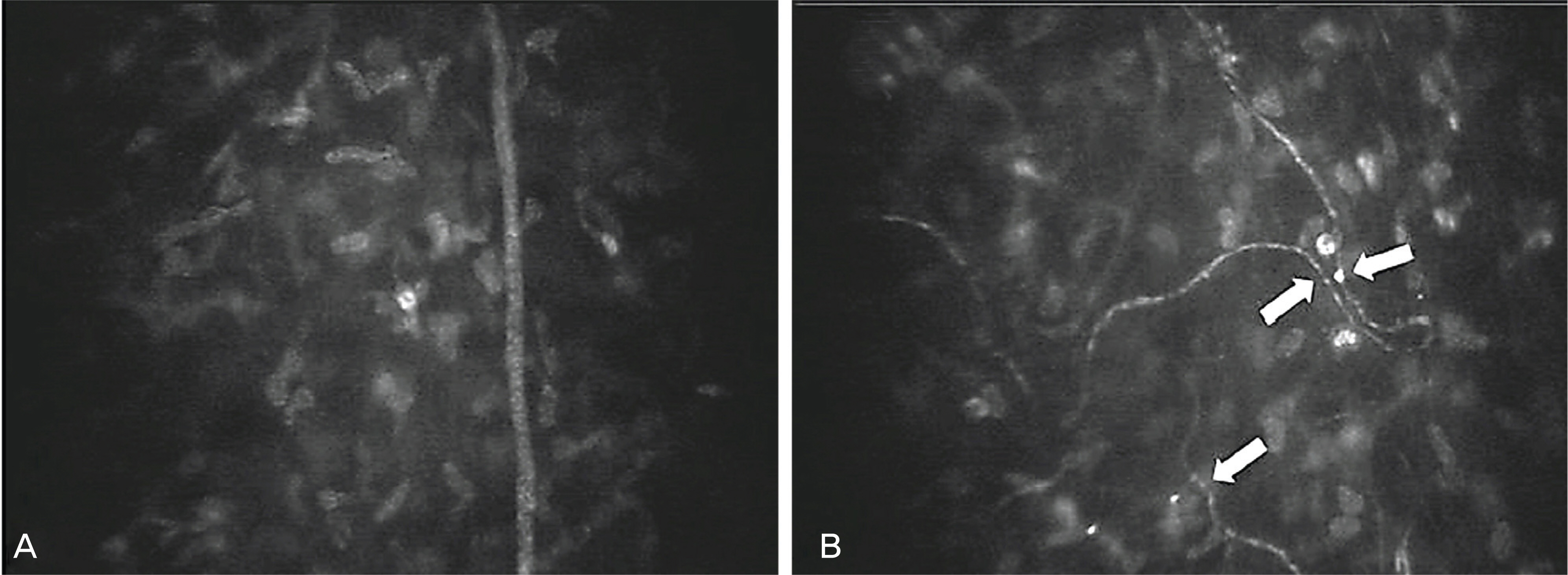
Figure 2. Corneal subbasal nerve plexus images of normal eye (A) and penetrating keratoplasty eye (B) digitalized directly from confocal microscopy through focusing scan (case 1). (A) normal subbasal nerve fibers. (B) abnormal subbasal nerve regeneration in the peripheral cornea after PKP. There were markedly increased nerve fiber tortuosity(arrows), beading, reduced branching pattern, and shorter nerve length.

Figure 3. Confocal microscopic images of 10 years after penetrating keratoplasty (case 2). All images are taken from a 60-year-old man (case 2). (A) normal anterior stroma. (B) activated keratocytes were apparent in anterior stroma. The hyper-reflectivity of activated keratocytes contributes to significant levels of corneal haze. Keratocytes with visible cytoplasm were prominent in the anterior part of the stroma. Reflective structures (arrows) represent the cytoplasmatic protrusions of an interconnected meshwork of keratocytes.
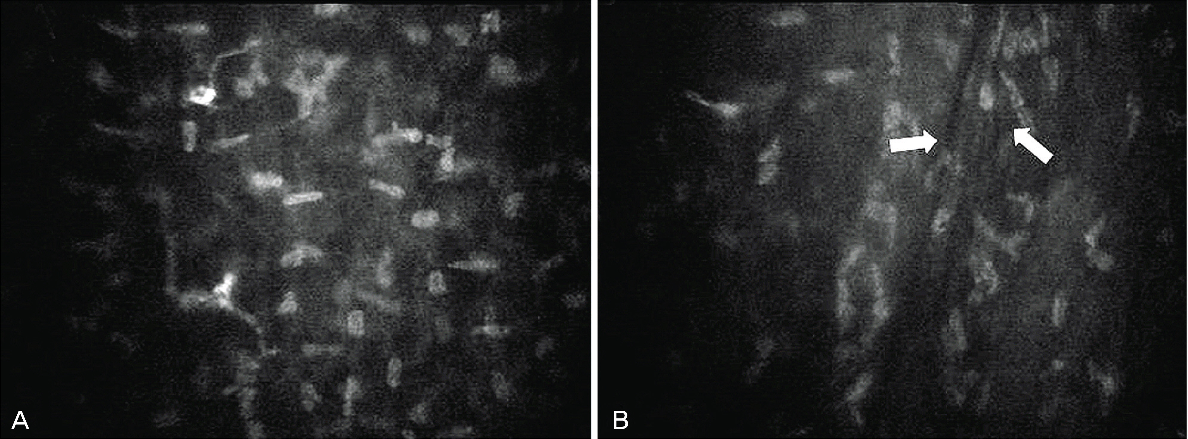
Figure 4. Confocal microscopic images of 10 years after penetrating keratoplasty (case 2). All images are taken from a 60-year-old man (case 2). (A) normal posterior stroma. (B) the keratocytes displayed irregular morphology and lower cell density than normal cornea. Because of corneal edema, hyporeflective folding lesions (arrows) were found on confocal microscopy.
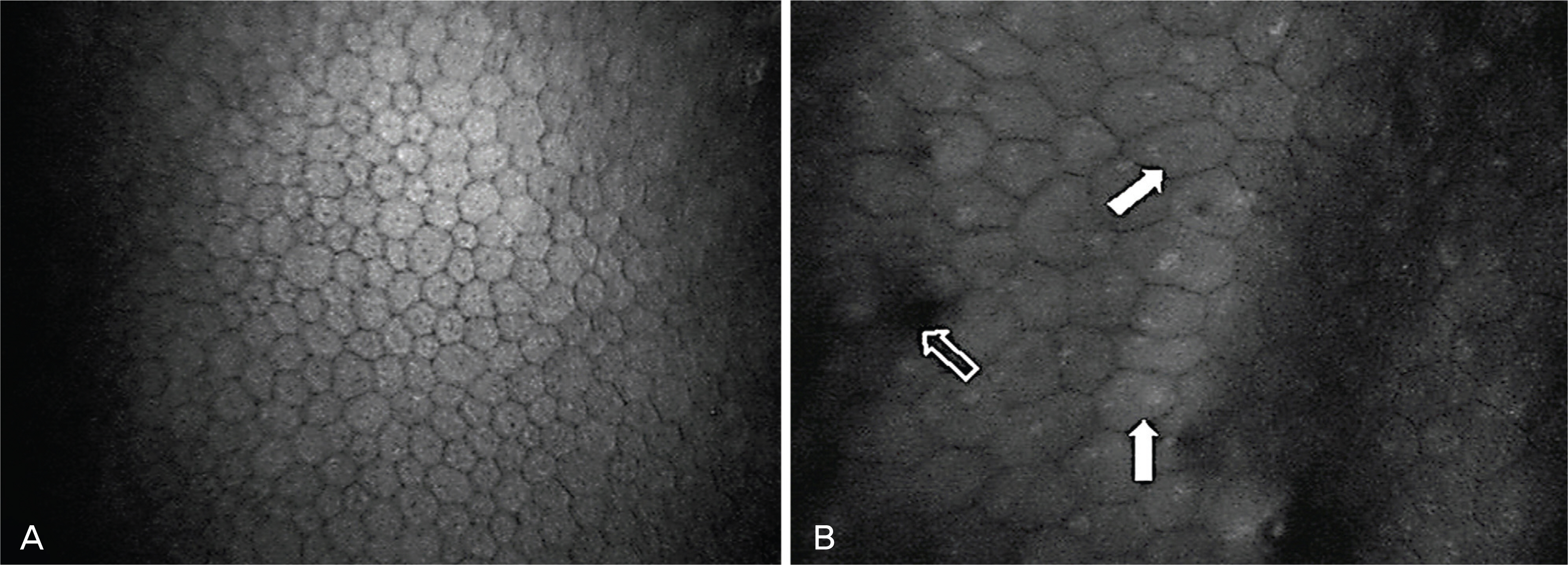
Figure 5. Corneal endothelial images of normal eye (A) and penetrating keratoplasty eye (B) digitalized directly from confocal microscopy through focusing scan in the same patient (case 1). (A) Normal endothelial cell density (2,998 cell/mm2). (B) Endothelial cell density and hexagonal cell density were decreased after penetrating keratoplasty (PKP). Multinucleated cells (white arrows) were seen in a case of PKP. The endothelial cell density was decreased to lower than normal endothelial cell density (2,057 cell/mm2). Corneal guttae (hollow arrow) appeared as roundish hyporeflective images.
Reference
-
References
1. Kim HS, Kim JH, Kim HM, Song JS. Comparison of Corneal Thickness Measured by Specular, US Pachymetry, and Orbscan in Post-PKP Eyes. J Korean Ophthalmol Soc. 2007; 48:245–50.2. Petroll WM, Cavanagh HD, Jester JV. Clinical confocal microscopy. Curr Opin Ophthalmol. 1998; 9:59–65.
Article3. Lee JS, Jong WH, Kim HK. Confocal Microscopic Corneal Findings in the Normal Rabbit and Human. J Korean Ophthalmol Soc. 2002; 43:739–44.4. Mustonen RK, McDonald MB, Srivannaboon S, et al. Normal human Corneal cell Populations Evaluated by In Vivo Scanning Slit Confocal Microscopy. Cornea. 1998; 17:485–92.
Article5. Chiou AG, Kaufman SC, Kaufman HE, Beuerman RW. Clinical Corneal Confocal Microscopy. Surv Ophthalmol. 2006; 51:482–500.
Article6. Szaflik JP, Kaminska A, Udziela M, Szaflik J. In vivo confocal microscopy of corneal grafts shortly after penetrating keratoplasty. Eur J Ophthalmol. 2007; 17:891–6.
Article7. Hollingsworth JG, Efron N, Tullo AB. A longitudinal case series investigating cellular changes to the transplanted cornea using confocal microscopy. Cont Lens Anterior Eye. 2006; 29:135–41.
Article8. Kohlhaas M. Corneal sensation after cataract and refractive surgery. J Cataract Refract Surg. 1998; 24:1399–409.
Article9. Neiderer RL, Perumal D, Sherwin T, McGhee CN. Corneal Innervation and cellular Changes after Corneal Transplantation: An In Vivo Confocal Microscopy Study. Invest Ophthalmol Vis Sci. 2007; 48:621–6.10. Moller-Pedersen T. Keratocyte reflectivity and corneal haze. Exp Eye Res. 2004; 78:553–60.11. Imre L, Resch M, Nagymihaly A. In vivo confocal corneal microscopy after keratoplasty. Ophthalmologe. 2005; 102:140–6.12. Bourne WM, Hodge DO, Nelson LR. Corenal endothelium five years after transplantation. Am J Ophthalmol. 1994; 118:185–96.13. Bertelmann E, Hartmann C, Scherer M, Rieck P. Outcome of rotational keratoplasty: comparison of endothelial cell loss in autografts vs allografts. Arch Ophthalmol. 2004; 122:1437–40.14. Ma DH, See LC, Chen JJ. Long-term observation of aqeous flare following penetrating keratoplasty. Cornea. 2003; 22:413–9.15. Ikebe H, Takamatsu T, Itoi M, Fujita S. Age-dependent changes in nuclear DNA content and cell size of presumably normal human corneal endothelium. Exp Eye Res. 1986; 43:251–8.
Article16. Patel DV, Phua YS, McGhee CN. Clinical and microstructural analysis of patients with hyperreflective corneal endothelial nuclei imaged by in vivo confocal microscopy. Exp Eye Res. 2006; 82:682–7.
Article
- Full Text Links
-
- Actions
-
Cited
- CITED
-
- Close
- Share
-
- Similar articles
-
- Clinical Evaluation of Full-thickness Deep Lamellar Keratoplasty
- A Case of Anterior Synechiolysis with Lamellar Corneal Dissection in Penetrating Keratoplasty
- Traumatic Wound Dehiscence after Penetrating Keratoplasty
- A Case of Macular Dystrophy of the Cornea
- Endothelial Cell Loss in Penetrating Keratoplasty
