J Korean Surg Soc.
2011 Sep;81(3):216-220. 10.4174/jkss.2011.81.3.216.
Intramural gallbladder hematoma mimicking gallbladder neoplasm in a 55-year-old male patient
- Affiliations
-
- 1Department of Internal Medicine, Eulji General Hospital, Eulji University School of Medicine, Seoul, Korea. sbk1026@eulji.ac.kr
- 2Department of Surgery, Eulji General Hospital, Eulji University School of Medicine, Seoul, Korea.
- 3Department of Pathology, Eulji General Hospital, Eulji University School of Medicine, Seoul, Korea.
- KMID: 2212200
- DOI: http://doi.org/10.4174/jkss.2011.81.3.216
Abstract
- Hemorrhage in the gallbladder (GB) is usually associated with cholecystitis, GB neoplasm, trauma, hemobilia, and cystic artery aneurysm. Our patient had not experienced any previous abdominal trauma, and GB hemorrhage was unlikely to result from cholecystitis or bleeding diathesis. A 55-year-old male was admitted because of right upper quadrant pain. Both prothrombin time and partial thromboplastin time were normal. Abdominal computed tomography, endoscopic ultrasound and magnetic resonance cholangiopancreatography were performed. Image studies revealed GB wall thickening and an intraluminal mass. Laparoscopic cholecystectomy was performed. Upon opening the GB postoperatively, a large amount of fresh blood and old blood clot was noted. The incidence of GB hematoma is very rare. GB hematoma should always be considered in the differential diagnosis of GB tumor. In such a situation, surgical intervention is needed for further patient evaluation and management. We present a rare case of intramural GB hematoma, of which we were unable to make a definitive diagnosis preoperatively.
Keyword
MeSH Terms
Figure
-
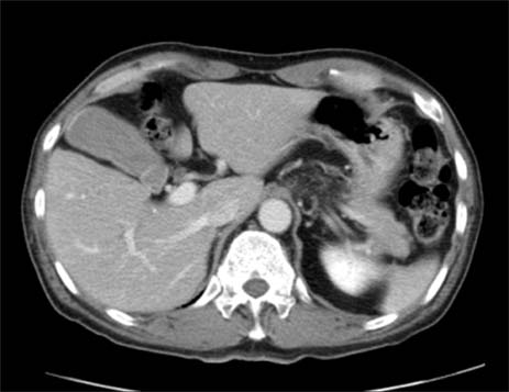
Fig. 1 Enhanced CT shows luminal high density in gallbladder.

Fig. 2 Endoscopic ultrasonography shows diffuse wall thickening of the gallbladder with a fixed echogenic polypoid mass (arrow) in the gallbladder lumen.

Fig. 3 MRCP shows (A) heterogenous multiple filling defects in gallbladder with diffuse gallbladder wall thickening (arrow) and (B) mild diffuse biliary tree dilatation without demonstrable obstructive lesion.

Fig. 4 Grossly the mucosal epithelium of gallbladder is covered by unwashed blood clots without any masslesion.

Fig. 5 Microscopic finding shows diffuse transmural hemorrhage with mucosal necrosis and infiltration of chronic inflammatory cells (H&E, ×40).
Reference
-
1. Tan SW, Lai SK, Ng KW, Chen P, Chen KH, Jiang CF. Intramural gallbladder hematoma mimicking gallbladder neoplasm in a 33-year-old male. J Chin Med Assoc. 2005. 68:146–149.2. Lai YC, Tarng DC. Hemorrhagic acalculous cholecystitis: an unusual location of uremic bleeding. J Chin Med Assoc. 2009. 72:484–487.3. Parekh J, Corvera CU. Hemorrhagic cholecystitis. Arch Surg. 2010. 145:202–204.4. Gore RM, Yaghmai V, Newmark GM, Berlin JW, Miller FH. Imaging benign and malignant disease of the gallbladder. Radiol Clin North Am. 2002. 40:1307–1323. vi5. Sandblom P. Hemorrhage into the biliary tract following trauma; traumatic hemobilia. Surgery. 1948. 24:571–586.6. Kwon TK, Jeon SH, Park HW, Jung WJ, Hwang JY, Park KS, et al. A case of intraluminal gallbladder hematoma after percutaneous liver biopsy. Korean J Hepatol. 2002. 8:486–489.7. Wibbenmeyer LA, Sharafuddin MJ, Wolverson MK, Heiberg EV, Wade TP, Shields JB. Sonographic diagnosis of unsuspected gallbladder cancer: imaging findings in comparison with benign gallbladder conditions. AJR Am J Roentgenol. 1995. 165:1169–1174.8. Gremmels JM, Kruskal JB, Parangi S, Kane RA. Hemorrhagic cholecystitis simulating gallbladder carcinoma. J Ultrasound Med. 2004. 23:993–995.9. Shimura T, Kojima T, Tsutsumi S, Yoshida T, Uchiumi H, Kuwano H. Gallbladder hematoma in a patient with hemophilia B, report of a case. Hepatogastroenterology. 2000. 47:939–941.10. Cho YU, Kim JY, Choi SK, Hur YS, Lee KY, Kim SJ, et al. A case of hemorrhagic gallbladder paraganglioma causing acute cholecystitis. Yonsei Med J. 2001. 42:352–356.
- Full Text Links
-
- Actions
-
Cited
- CITED
-
- Close
- Share
-
- Similar articles
-
- Intramural Hypoattenuated Nodules in Thickened Wall of the Gallbladder: CT Features According to Their Primary Causes
- Non-Operative Management of Traumatic Gallbladder Bleeding with Cystic Artery Injury: A Case Report
- Intramural Duodenal Hematoma Caused by Acute Gallstone Pancreatitis
- Strangulation of the Floating Gallbladder by the Lesser Omentum
- A Case of Metastatic Renal Cell Carcinoma to the Gallbladder
