Myoepithelial carcinoma with contralateral invasive micropapillary carcinoma of the breast
- Affiliations
-
- 1Department of Pathology, Keimyung University School of Medicine, Daegu, Korea. pathol72@dsmc.or.kr
- 2Department of Pathology, Yeungnam University College of Medicine, Daegu, Korea.
- 3Department of Surgery, Keimyung University School of Medicine, Daegu, Korea.
- KMID: 2212199
- DOI: http://doi.org/10.4174/jkss.2011.81.3.211
Abstract
- Adenomyoepithelioma (AME) is a rare benign tumor composed of myoepithelial cells (MECs) which are located beneath the epithelial cells of exocrine glands, especially in breast and salivary glands. These tumor cells show biphasic proliferation of epithelial and MECs. Malignant AME is characterized by distant metastasis, local recurrence, cytologic atypia, high mitotic activity and infiltrating tumor margins. A 51-year-old woman presented with an 8 months growth in the left breast. She underwent core-needle biopsy and consecutively mammotome assisted biopsy at a local clinic. After resection, she complained about re-growing remnant lesion and a newly developed solid mass in the right breast. Finally, the remnant mass in the left breast was diagnosed with myoepithelial carcinoma. Concurrently, contralateral breast mass was diagnosed with invasive micropapillary carcinoma. Herein we report an unusual case of synchronous myoepithelial carcinoma and invasive micropapillary carcinoma of the breast with a review of literatures.
MeSH Terms
Figure
-
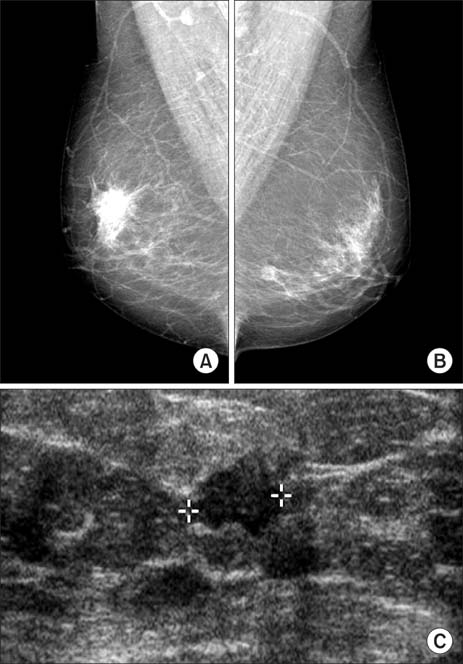
Fig. 1 (A) Right mammogram showed a spiculated and heterogenous nodule without a focal lesion or microcalcification. (B) Left mammogram demonstrated a lobulated and iso-density nodule. (C) Preoperative ultrasonogram of the left breast revealed a 1.0 cm sized inhomogeneous, irregular marginated and hypoechoic mass.
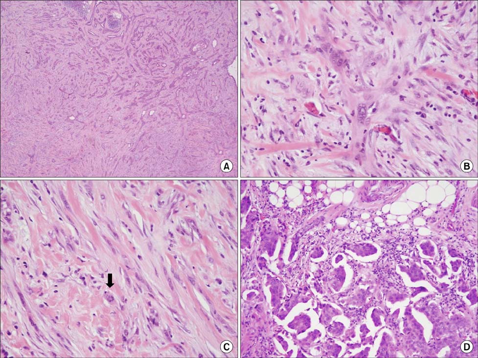
Fig. 2 Microscopic findings of the left breast mass (A-C) and the right breast mass (D). (A) The central lesion of the left breast mass demonstrated adenomyoepithelioma with atypical spindle cells at the left lower corner (H&E, ×100). (B) The atypical spindle tumor cells showed round to spindle, hyperchromatic and pleomorphic nuclei with relatively abundant eosinophilic cytoplasm (H&E, ×400). (C) Atypical mitotic figure (arrow) was found (H&E, ×400). (D) Invasive micropapillary carcinoma was identified in the right breast (H&E, ×200).

Fig. 3 Myoepithelial carcinoma revealed diffuse strong reactivity for smooth muscle actin (A, immunohistochemical stain, ×200), calponin (B, immunohistochemical stain, ×200), p63 (C, immunohistochemical stain, ×200) and pan-cytokeratin (D, immunohistochemical stain, ×200).
Reference
-
1. Tavassoli FA. Myoepithelial lesions of the breast. Myoepitheliosis, adenomyoepithelioma, and myoepithelial carcinoma. Am J Surg Pathol. 1991. 15:554–568.2. Sarkar K, Kallenbach E. Myoepithelial cells in carcinoma of human breast. Am J Pathol. 1966. 49:301–307.3. Hamperl H. The myothelia (myoepithelial cells). Normal state; regressive changes; hyperplasia; tumors. Curr Top Pathol. 1970. 53:161–220.4. Tavassoli F, Devilee P. Pathology and genetics of tumours of the breast and female genital organs. 2003. Geneva: World Health Organization.5. Loose JH, Patchefsky AS, Hollander IJ, Lavin LS, Cooper HS, Katz SM. Adenomyoepithelioma of the breast. A spectrum of biologic behavior. Am J Surg Pathol. 1992. 16:868–876.6. Ahmed AA, Heller DS. Malignant adenomyoepithelioma of the breast with malignant proliferation of epithelial and myoepithelial elements: a case report and review of the literature. Arch Pathol Lab Med. 2000. 124:632–636.7. Tavassoli FA. Pathology of the breast. 1999. 2nd ed. Stamford: Appleton & Lange.8. Han B, Mori I, Nakamura M, Wang X, Ozaki T, Nakamura Y, et al. Myoepithelial carcinoma arising in an adenomyoepithelioma of the breast: case report with immunohistochemical and mutational analysis. Pathol Int. 2006. 56:211–216.9. Jones C, Tooze R, Lakhani SR. Malignant adenomyoepithelioma of the breast metastasizing to the liver. Virchows Arch. 2003. 442:504–506.10. Angèle S, Jones C, Reis Filho JS, Fulford LG, Treilleux I, Lakhani SR, et al. Expression of ATM, p53, and the MRE11-Rad50-NBS1 complex in myoepithelial cells from benign and malignant proliferations of the breast. J Clin Pathol. 2004. 57:1179–1184.
- Full Text Links
-
- Actions
-
Cited
- CITED
-
- Close
- Share
-
- Similar articles
-
- Invasive Micropapillary Carcinoma in Axillary Ectopic Breast and Synchronous Ductal Carcinoma In Situ in the Contralateral Breast
- Invasive Micropapillary Carcinoma of the Breast: A clinicopathologic study of 16 cases
- Invasive Micropapillary Carcinoma in Breast Presented as Hyperechoic Mass with Coarse Macrocalcifications: A Case Report
- Fine Needle Aspiration Cytology of Invasive Micropapillary Carcinoma of the Breast
- Prognostic Significance of a Micropapillary Pattern in Pure Mucinous Carcinoma of the Breast: Comparative Analysis with Micropapillary Carcinoma
