Anatomy and Physiology of Lumbar Spine
- Affiliations
-
- 1Department of Orthopaedic Surgery, College of Medicine, Konyang University, Taejeon, Korea.
- KMID: 2209745
- DOI: http://doi.org/10.4184/jkss.2001.8.3.264
Abstract
- The spinal column is separated into the 7 cervical vertebra, the 12 thoracic vertebra, the 5 lumbar vertebra, the 5 sacral vertebra and the 4 coccygeal vertebra. The cervical, thoracic and lumbar vertebra are named as the movable vertebra and the sacral and coccygeal vertebra are named as the fixed vertebra. The lumbar spine includes five large vertebra situated between the rela-tively immobile rib cage and the pelvis. A typical lumbar vertebra has 2 main structures which are vertebral body and vertebral arch. The vertebral body is the anterior portion of a vertebra and the vertebral arch is the posterior portion of it and surrounds the vertebral foramen. In contrast to thoracic vertebra, lumbar vertebra has a wide disc space, sagittally oriented facets, and suf-ficient space between its lamina to permit a considerable range of motion. This report will explains some important normal anatomic features of the lumbar spine and sacrum including with their musclatures and neurovascular structures.
Keyword
Figure
-

Fig. 1. Lumbar vertebra(Lateral view).1; Superior articular process. 2; Superior articular surface. 3; Vertebral body. 4; Inferior articular process. 5; Spinous process.6; Pedicle. 7; Transverse process. 8; Mammillary process.

Fig. 2. Fifth lumbar vertebra(Cranial view).1; Superior articular process. 2; Superior articular surface. 3; Vertebral arch(lamina). 4; Spinous process. 5; Vertebral foramen. 6; Inferior articular process. 7; Transverse process. 8; Pedicle. 9; Vertebral body.
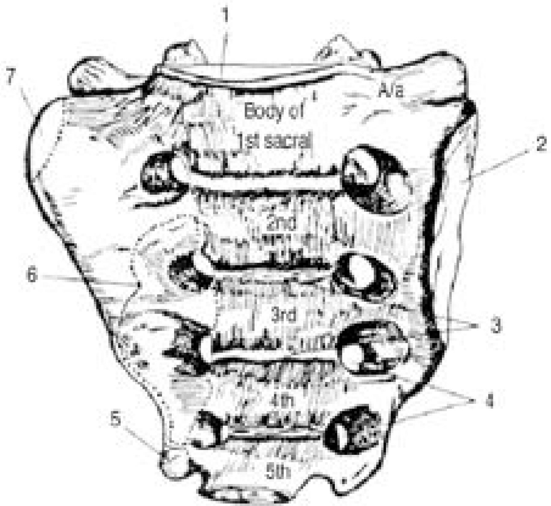
Fig. 3. Pelvic surface of the sacrum.1; Superior terminal surface. 2; Auricular surface of sacrum. 3; Transverse lines(ridges). 4; Pelvic sacral foramen. 5; Inferior lateral angle. 6; Piriformis. 7; Iliacus.

Fig. 4. Dorsal surface of the sacrum.1; Superior articular process. 2; Posterior sacral foramen. 3; Inferior lateral angle. 4; Sacral cornua. 5; Apex of sacrum. 6; Sacral hiatus. 7; Gluteus maximus. 8; Erector spinae. 9; Sacral tuberosity. 10; Multifidus.

Fig. 5. Lumbar vertebra(Midsagittal view).1; Anterior longitudinal ligament. 2; Vertebral body. 3; Posteior longitudinal ligament. 4; Pedicle. 5; Vertebral arch(lamina). 6; Superior articular process. 7; Ligamentum flavum. 8; Supraspinous ligament. 9; Spinous process. 10; Interspinous ligament. 11; Joint capsule. 12; Basivertebral vein. 13; Intervertebral disc.

Fig. 6. Posterior longitudinal ligament of Lumbar vertebra.1; Pedicle. 2; Intervertebral disc. 3; Posterior longitudinal ligament.
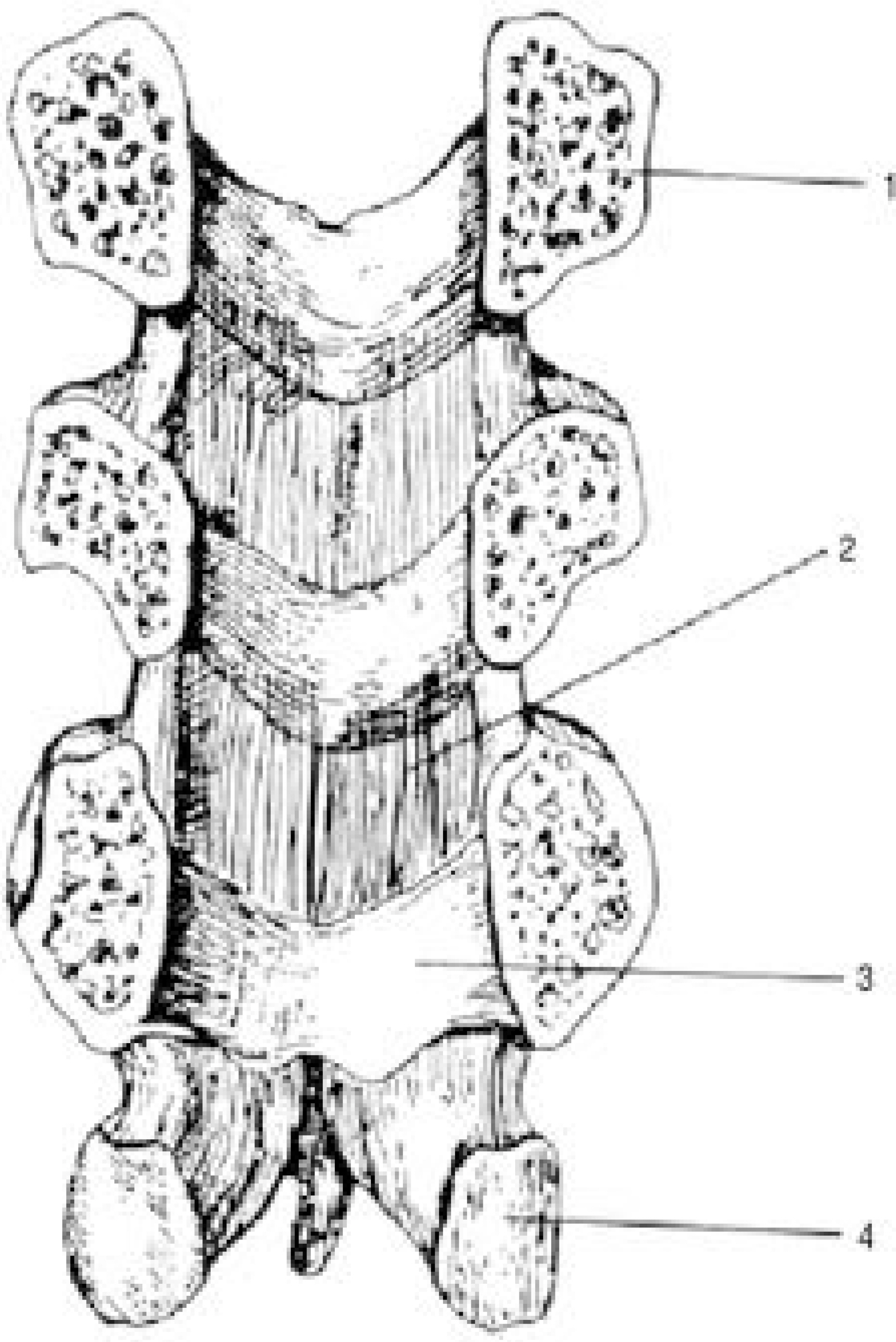
Fig. 7. Ligamentum flavum of Lumbar Vertebra.1; Pedicle. 2; Ligamentum flavum. 3; Vertebral arch(lamina).4; Inferior articular process.
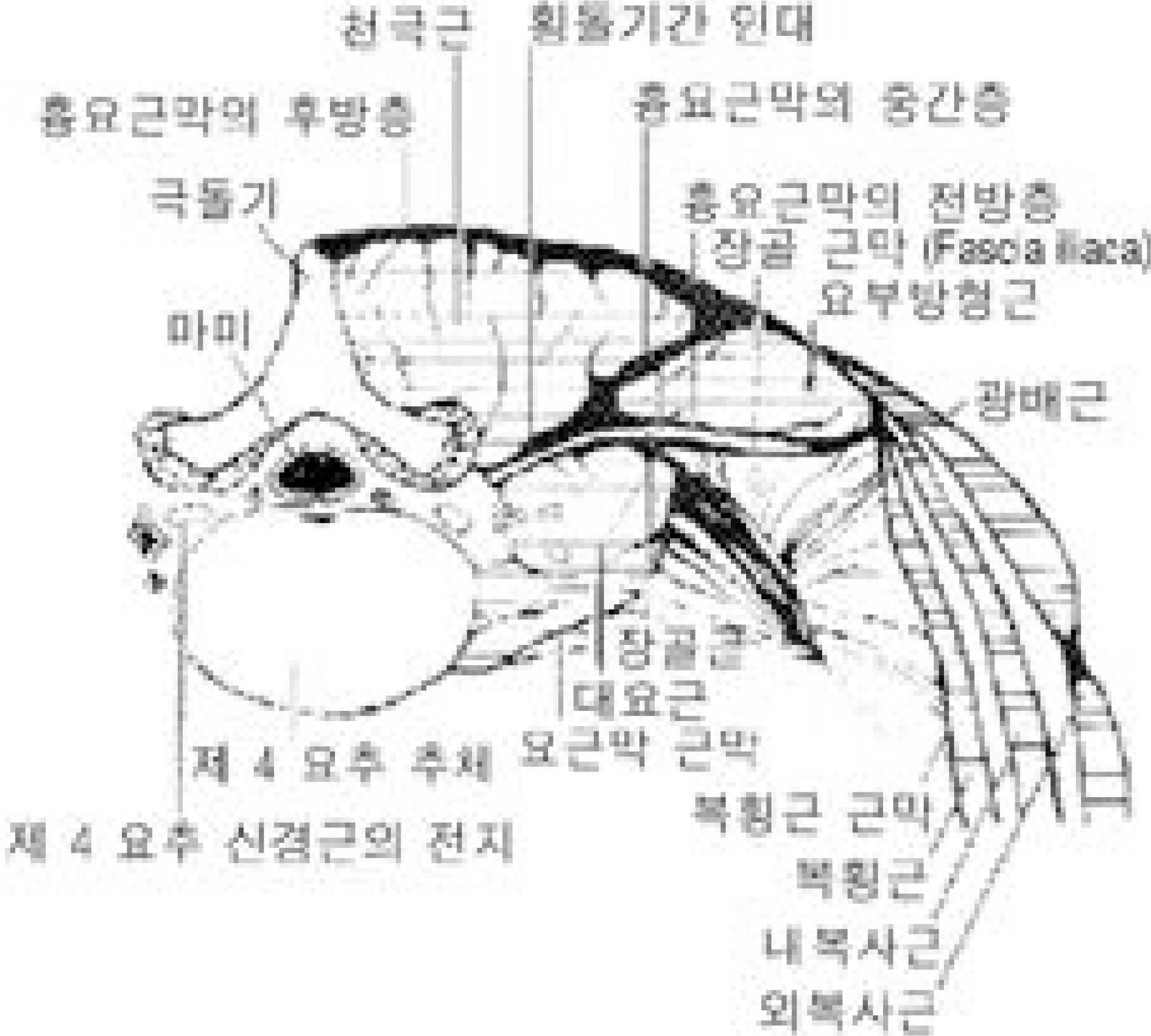
Fig. 8. Cross section of Back, 4th lumbar vertebra.
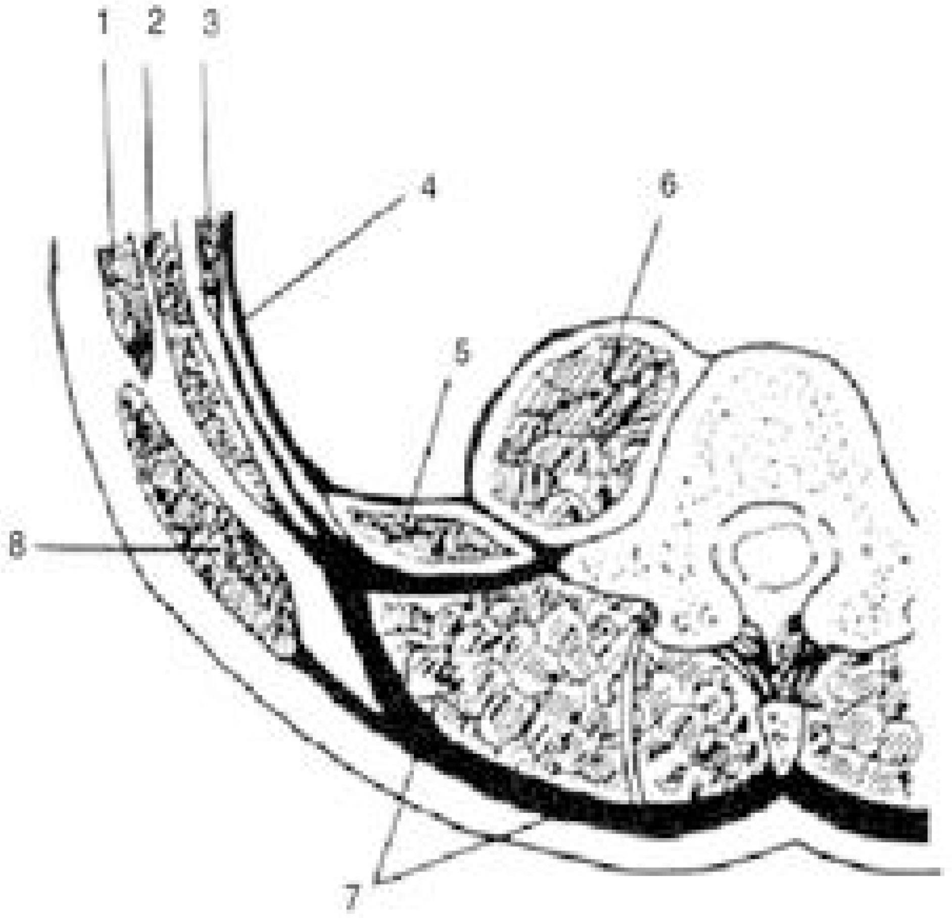
Fig. 9. Thoracolumbar fascia.1; External oblique muscle. 2; Internal oblique muscle. 3; Transversus abdominis muscle. 4; Transversalis fascia. 5; Quadratus lumborum muscle. 6; Psoas major muscle. 7; Thoracolumbar fascia. 8; Latissimus dorsi muscle.
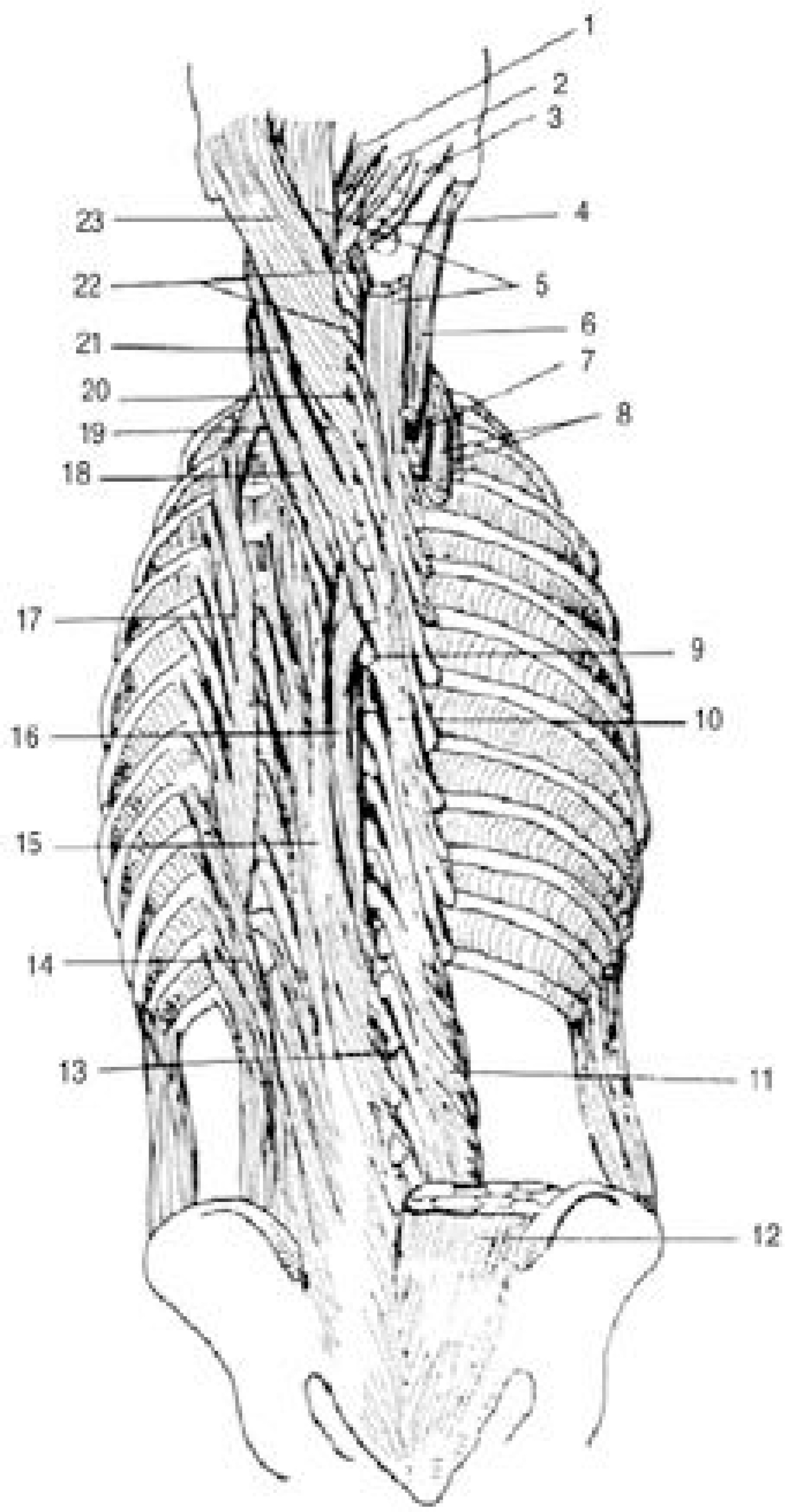
Fig. 10. Muscles of the vertebral column.1; Rectus capitis posterior minor. 2; Rectus capitis posterior major. 3; Obliquus capitis superior. 4; Obliquus capitis inferior. 5; Semispinalis capitis. 6; Logissimus capitis. 7; Longissimus cervicis. 8; Iliocostalis cervicis and thoracis. 9; 6th thoracic vertebra. 10; Semispinalis thoracis. 11; Multifidus. 12; Erector spinae. 13; 1st lumbar vertebra. 14; Iliocostalis lumborum. 15; Longissimus thoracis. 16; Spinalis thoracis. 17; Iliocostalis thoracis. 18; Longissimus cervicis. 19; Iliocostalis cervicis. 20; 7th cervical vertebra. 21; Splenius cervicis. 22; Semispinalis cervicis. 23; Splenius capitis.
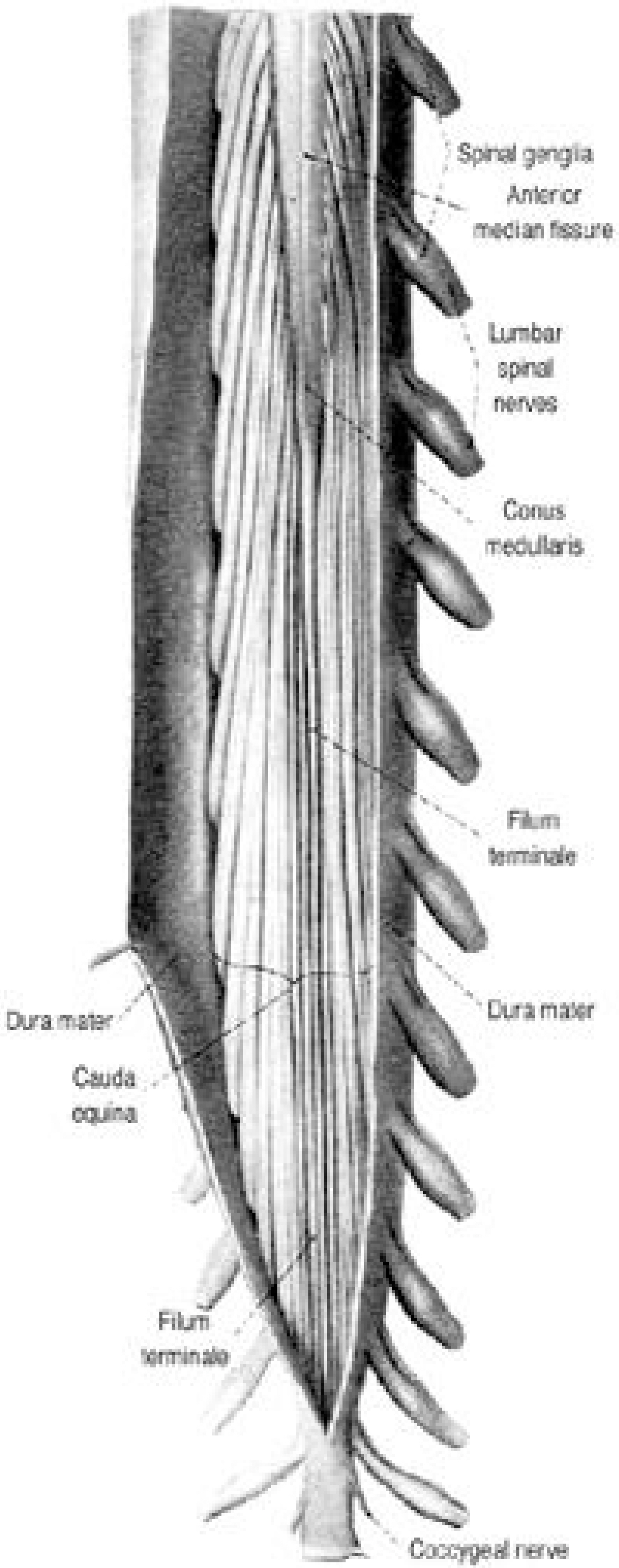
Fig. 11. Conus Medullaris and Cauda Equina(Ventral)

Fig. 12. Internal and external vertebral venous plexus.
Reference
-
1). Adams MA, Hutton WC. The effect of posture on the role of the apophyseal joints in resisting intervertebral compressive forces. J Bone Joint Surg. 62B:358–362. 1980.2). Basmajian JV. Grant's method of anatomy. 10th ed.Baltimore: Williams & Wilkins;p. 325–326. 1980.3). Batson OV. The function of the vertebral veins and their role in the spread of metastases. Ann Surg. 112:138–149. 1940.
Article4). Clement CD. Gray's anatomy. 30th ed.Philadelphia: Lea & Febiger;p. 119–147. 341-366, 456-475. 1985.5). Coventry MB, Ghormley RK, Kernohan JW. The intervertebral disc: Its microscopic anatomy and pathology. Part I. Anatomy, development and physiology. J Bone Joint Surg. 27:105–112. 1945.6). Elliot HC. Cross-sectional diameters and areas of the human spinal cord. Ana Rec. 93:287. 1943; (cited from Hollinshead WH: Anatomy for Surgeons: vol. 3, The back and limbs, 3rd ed. Philadelphia, Harper & Row, 1982).7). Gregersen GG, Lucas DB. An in vivo study of the axial rotation of the human thoracolumbar spine. J Bone Joint Surg. 49-A:247–262. 1967.
Article8). Haldeman S. Spinal clinical neurophysiology.(in Cau -then JC eds. Lumbar Spine Surgery. 2nd ed.Baltimore: Williams & Wilkins;p. 128–143. 1988. .).9). Kim NH, Lee HM. Spinal surgery. Seoul: Eui-Hak Publishing Co;p. 1–31. 1998.
Article10). Pauly JE. An electromyographic analysis of certain movements and exercises. I. Some deep muscles of the back. Anat Rec. 155:223–234. 1966.
Article11). Sadler TW. Langman's Medical Embryology. 6th ed.Baltimore: Williams & Wilkins;p. 139–152. 1990.12). Sensenig EC. The early development of the human vertebral column. Vontr Embryol Carneg Instn. 33:21–41. 1949.13). Steel HH. Anatomical and mechanical considerations of the atlantoaxial articulations. In proceedings of the American Orthopaedic Association. J Bone Joint Surg. 50A:1481–1482. 1968.14). Suk SI. Spinal surgery. Seoul: Newest Medical Journal;p. 15–29. 1997.
Article15). Suk SI, Lee CK, Kim WJ, Chung YJ, Song KY, Park YB. Segmental pedicle screw fixation in the treatment of thoracic idiopathic scoliosis. J of Korean Orthop. 30:49–58. 1995.
Article
- Full Text Links
-
- Actions
-
Cited
- CITED
-
- Close
- Share
-
- Similar articles
-
- The Change of Lumbar Lordosis after Pedicular Screw Fixation of the Degenerative Lumbar Spine
- New signs in observation of posterior arches of lumbar vertebrae
- Ultrasonography for lumbar neuraxial block
- Relative Contribution of Upper and Lower Lumbar Spinal Segments to Flexion/Extension: Comparison between Normal Spines and Spines with Disc Disease in Asian Patients
- Letter to the Editor: Analysis of Functional and Radiological Outcome Following Lumbar Decompression without Fusion in Patients with Degenerative Lumbar Scoliosis
