Ossification of Posterior Longitudinal Ligament
- Affiliations
-
- 1Spine Center, Department of Orthopaedic Surgery, Sun General Hospital, Daejeon, Korea. chspine@korea.com
- KMID: 2209636
- DOI: http://doi.org/10.4184/jkss.2006.13.3.153
Abstract
- No abstract available.
Figure
-
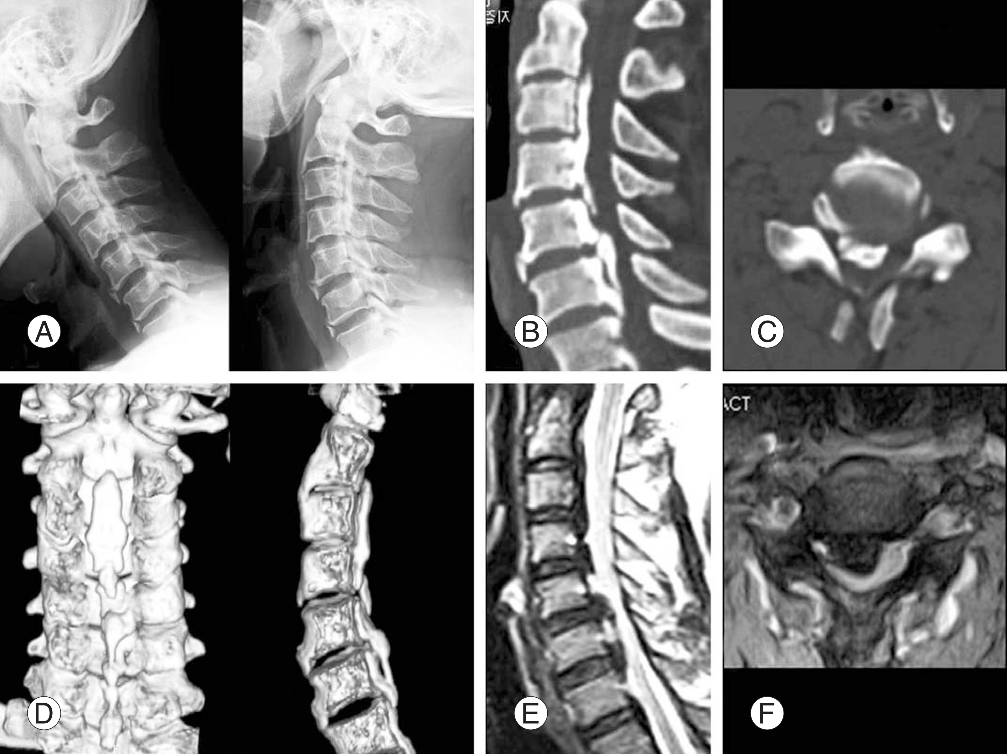
Fig. 1. Diagnostic imagings of cervical OPLL. (A) Flexion / Extenion dynamic radiograms show no definite detetectable motion at the continous portion of OPLL. OPLL is frequently overlooked on plain radiograms because of superimposed bony structures such as facets and laminae. (B) Sagittal reconstruction CT scan shows a long strip of ossification posterior to the C2-C4 verte-bral bodies and mixed configuration at C5-7. (C) Axial CT shows ossified mass with a hole inside enchroaching spinal canal. (D) Three dimensional reconstruction CT scans show coronal and sagittal configuration of OPLL. (E) Sagittal T2-weighted MRI shows bandlike low or no signal intensity of ossified mass compressing spinal cord and small linear high-intensity area (fatty marrow) posterior to C6 and intramedullary bright signal posterior to C5-6. (F) Axial T-weighted MRI shows fuge ossified mass compressing spinal cord.
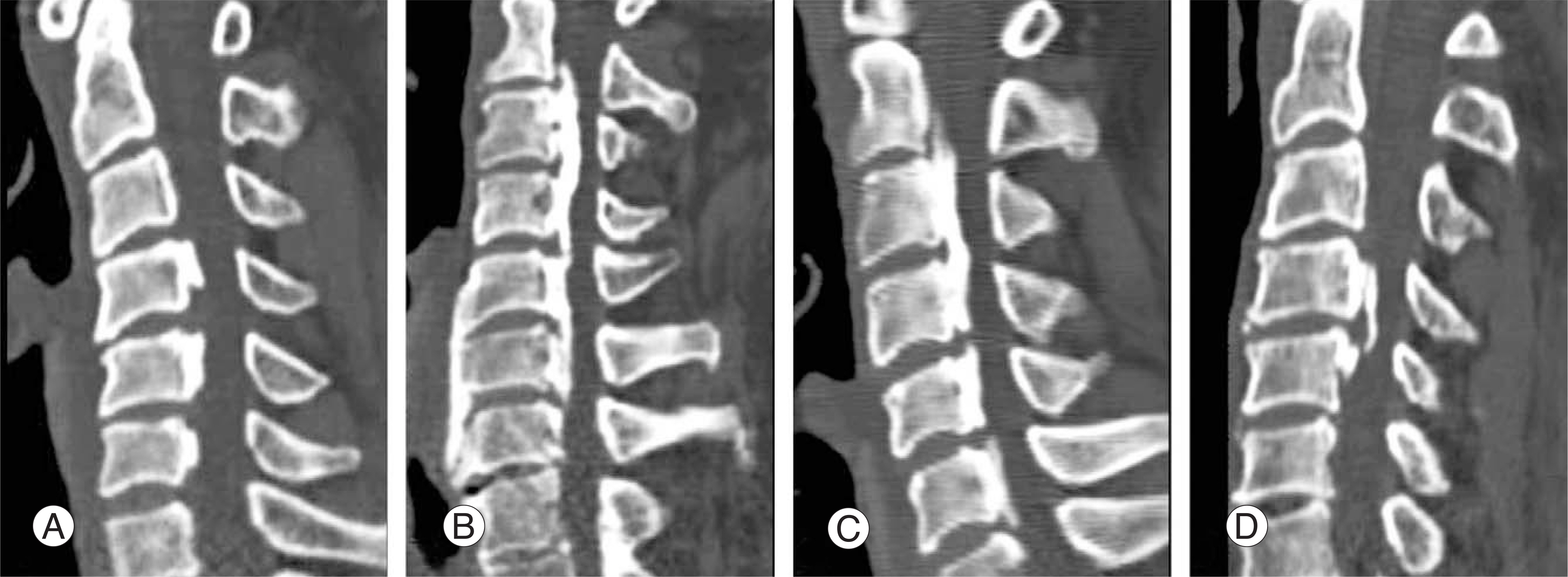
Fig. 2. Classification of OPLL by Investigation Committee of Japan. (A) Segmental type (B) Continuous type (C) Mixed type (D) Other (localized) type

Fig. 3. OPLL in Evolution. Axial T2-weighted MRI (A) and CT scan (B) show OEV-like mass compressing spinal cord. OEV mass, especially at C5-6, shows inhomogeneity of the mixed signals reflecting hypertrophied ligamentous and calcified or ossified contents.
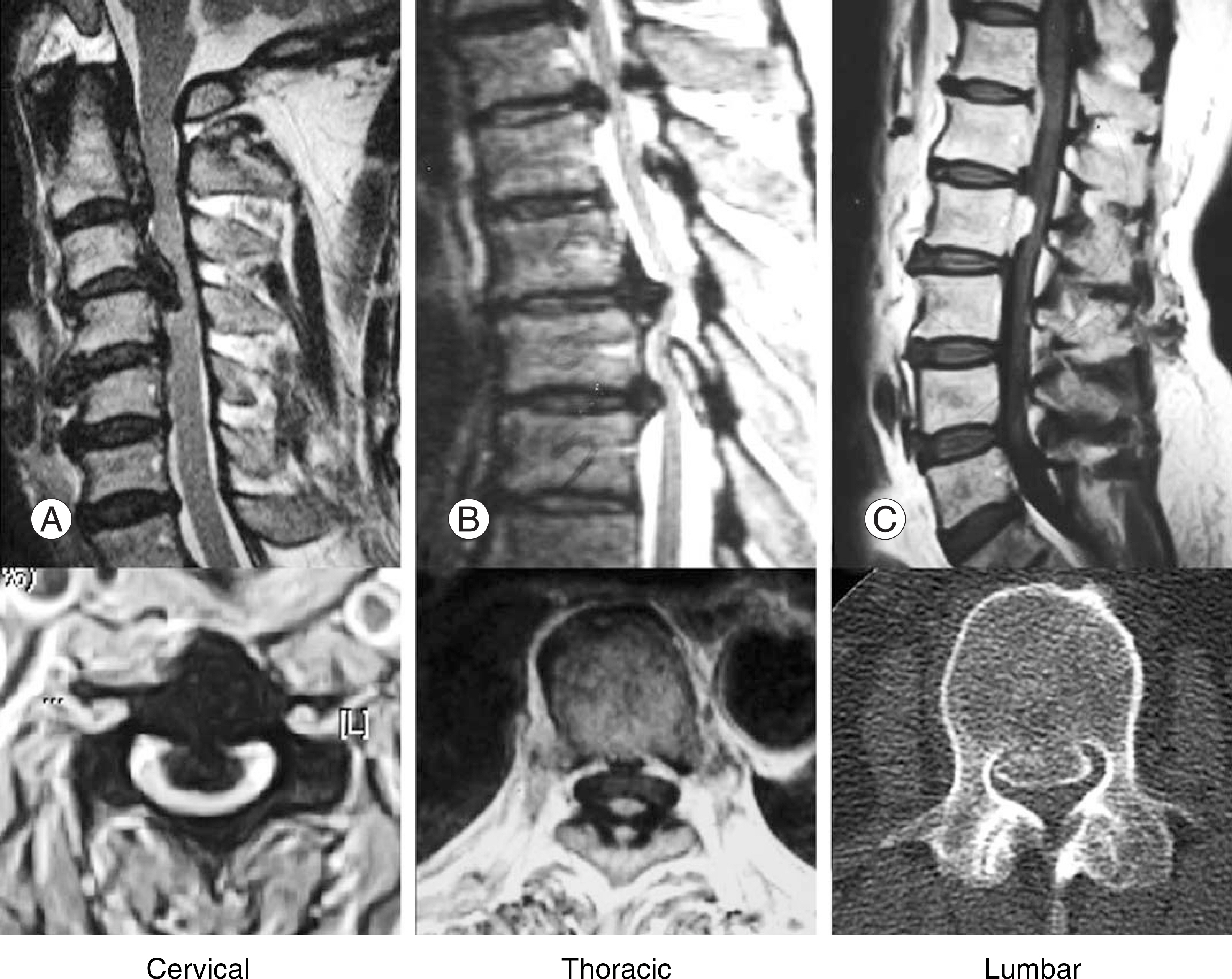
Fig. 4. A 54-year-old female with OPLL of cervical (A), Thoracic (B) and lumbar spine (C) and ossification of yellow ligament (OYL) of Thoracic spine (B).
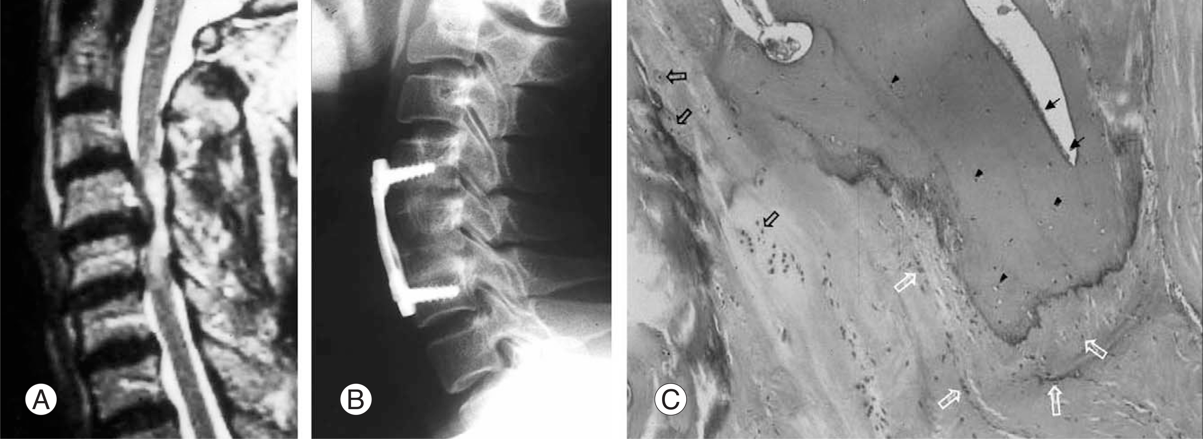
Fig. 5. Anterior decompressive surgery. (A) Sagittal T2-weighted MRI shows localized type of OPLL compressing spinal cord at C4-5. (B) Post-op. lateral radiogram shows partial corpectomy of C4,5 and iliac bone graft and plate fixation. (C) Hematoxylin-eosin staining of the OPLL specimen achieved from same patient showing osteoblast (black arrows) and osteocytes (black arrowheads) in ossified areas, chondrocytes (black hollow arrows) in cartilaginous areas, and fibroblasts (white hollow arrows) in unossified areas (×40).

Fig. 6. A 67-year-old male with cervical OPLL complained of both hand clumsiness and walking difficulty. (A) Pre-op. T2-weighted MRI show marked cord compression with intramedullary high signal intensity at C4-5. (B) Post-op. T2-weighted MRI show expansion of spinal cord and recovery of subarachnoid space and still show intramedullary high signal intensity at the previ-ous maximum cord compression area.

Fig. 7. Posterior migration of spinal cord is more effective in lordotic cervical curve (A), however, local dural expansion effect can be expected even in straight (B) and kyphotic cervical curve (C).
Reference
-
01). Inaba H., Nakae K., Kurokawa T. Statistical analysis for the national survey of ossification of the posterior longitudinal ligaments (in Japanese). In: Investigation committee 1976 report on the ossification of the spinal ligaments of the Japanese ministry of Public Health and Welfare, Tokyo. 1977. 29–39.02). Tsuyama N. Ossification of the posterior longitudinal ligament of the spine. Clin Orthop. 1984. 184:71–84.
Article03). Key CA. Paraplegia depending on disease of the ligaments of the spine. Guy' s Hosp Rep. 1938. 3:17–34.04). Tsukimoto H. A case report: autopsy of syndrome of compression of spinal cord owing to ossification within spinal canal of cervical spine(in Japanese). Nippon Geka Hokan. 1960. 29:1003–1007.05). Yamaura I., Isobe Y., Kamikozuru M, et al. Evaluation of surgical treatment of the posterior longitudinal ligament in the cervical spine; anterior decompression (in Japanese). Seikeigeka. 1976. 27:87–95.06). Yamaura I. Anterior approach (anterior floating method) and its surgical results for cervical myelopathy caused by ossification of the posterior longitudinal ligament (OPLL). J West Pac Orthop Assoc. 1990. 17:47–55.07). Hirabayashi K. Expansive open-door laminoplasty for cervical spondylotic myelopathy (in Japanese). Geka (surgery). 1978. 32:1159–1163.08). Hirabayashi K., Miyakawa J., Satomi K., Maruyama T., Wakano K. Operative results and postoperative progression of ossification among patients with ossification of cervical posterior longitudinal ligament. Spine. 1981. 6:354–364.
Article09). Hirabayashi K., Satomi K. Operative procedure and results of expansive open-door laminoplasty. Spine. 1988. 13:870–876.
Article10). Kurokawa T. Enlargement of spinal canal by the sagittal splitting of spinous processes (in Japanese). Bessatsu Seikeigeka. 1982. 2:234–240.11). Itoh T., Tsuji H. Technical improvements and results of laminoplasty for compressive myelopathy in the cervical spine. Spine. 1985. 10:729–736.
Article12). Kawai S., Sunago K., Doi K., Sakai M., Toguchi T. Cervical laminoplasty (Hattori' s method). Spine. 1988. 13:1245–1250.13). Tomita K., Nomura S., Umeda S., Baba K. Cervical laminoplasty to enlarge the spinal canal in multilevel ossification of the posterior longitudinal ligament in myelopathy. Arch Orthop Trauma Surg. 1988. 107:148–153.14). Matsunaga S., Sakou T., Taketomi , et al. Effects of strain distribution in the intervertebral discs on the progression of ossification of the posterior longitudinal ligaments. Spine. 1996. 21:184–189.
Article15). Matsunaga S., Kukita M., Hayashi K, et al. Pathogenesis of myelopathy in patients with ossification of the posterior longitudinal ligament. J Neurosurg Spine. 2002. 2:168–172.
Article16). Taketomi E. Progression of the posterior longitudinal ligament in the cervical spine. J Jpn Spine Res Soc. 1997. 8:359–366.17). Chiba K., Yamamoto I., Hirabayashi H, et al. Multicen-ter study investigating the postoperative progression of ossification of the posterior longitudinal ligament in the cervical spine; a new computer-assisted measurement. J Neurosurg Spine. 2005. 3:17–23.
Article18). Epstein N. Ossification of the posterior longitudinal liga-ment; Diagnosis and surgical management. Neurosurg Q. 1992. 2:223–241.19). Chang H., Bahk WJ., An JG., Choi KH. Pre and postoperative evaluation of cervical myelopathy using MR imaging. J Korean Soc Spine Surg. 1994. 1:326–336.20). Matsunaga S., Sakou T., Taketomi , et al. The natural course of myelopathy caused by ossification of the posterior longitudinal ligaments in the cervical spine. Clin Orthop. 1994. 305:168–177.21). Masunaga S., Sakou T., Hayashi K., Ishidou Y., Hirotsu M., Komiya S. Trauma-induced myelopathy in patients with ossification of the posterior longitudinal ligament. J Neurosurg Spine. 2002. 2:172–175.22). Ogawa Y., Chiba K., Matsumoto M, et al. Long term results after expansive open-door laminoplasty for the segmental-type of ossification of the posterior longitudinal ligament of the cervical spine: a comparison with non-seg-mental type lesion. J Neurosurg Spine. 2005. 3:198–204.23). Mizuno J., Nakagawa H., Matsno N., Song J. Dural ossi-fication associated with cervical ossification of posterior longitudinal ligament: frequency of dural ossification and compression of neuroimaging modalities in ability to iden-tify the disease. J Neurosurg Spine. 2005. 2:425–430.24). Epstein N. Circumferential cervical surgery for ossification of posterior longitudinal ligament, A multianalytic outcome study. Spine. 2004. 29:1340–1345.25). Kawai S., Sunago K., Doi K., Sakai M., Taguchi T. Cervical laminoplasty (Hattori' s method). Spine. 1988. 13:1245–1250.26). Iwasaki M., Kawaguchi Y., Kimmura T., Yonenobu K. Long term result of expansive laminoplasty for ossification of the posterior longitudinal ligament of the cervical spine: more than 10 years follow up. J Neurosurg Spine. 2002. 2:180–189.27). Yonenobu K., Okada K., Fuji T, et al. Causes of neuro-logic deterioration following surgical treatment of cervical myelopathy. Spine. 1991. 11:818–823.
Article28). Tsuzuki N., Abe R., Saiki K, et al. Paralysis of the arm after posterior decompression of the spinal cord II; Analy-ses of Clinical Findings. Eur Spine J. 1993. 2:197–202.29). Tsuzuki N., Abe R., Saiki K, et al. Extradural tethering effect as one mechanism of radiculopathy complicating posterior decompression of the cervical spinal cord. spine. 1996. 21:203–211.
Article30). Dai L., Ni B., Yuan W, et al. Radiculopathy after laminoplasty for cervical compressive myelopathy. J Bone Joint Surg Br. 1998. 80:846–849.31). Chiba K., Toyama Y., Matsumotor M, et al. Segmental motor paralysis after expansive open door laminoplasty. Spine. 2002. 27:2108–2115.32). Sakura H., Hosomo N., Mukai Y., Ishii T., Yoshikawa H. C5 palsy after decompression surgery for cervical myelopa-thy, Review of the literature. Spine. 2003. 28:2447–2451.33). Seiki A., Takeshita K., Kawaguchi ., Nakajima S., Akune T., Nakamura K. Postoperative expansion of intramedullary high intensity areas on T2-weighted magnetic resonance imaging after cervical laminoplasty. Spine. 2004. 29:1478–1482.
- Full Text Links
-
- Actions
-
Cited
- CITED
-
- Close
- Share
-
- Similar articles
-
- Ossification of the Posterior Longitudinal Ligament: 2 cases report
- Ossification of posterior longitudinal ligament of the cervical spine in Korean
- Does Ossification of the Posterior Longitudinal Ligament Progress after Fusion?
- Anterior Decompression and Interbody Fusion for Ossification of the Posterior Longitudinal Ligament of the Cervical Spine: Case Report
- Genetics of Ossification of the Posterior Longitudinal Ligament of the Spine: A Mini Review
