J Korean Soc Radiol.
2010 Feb;62(2):159-162. 10.3348/jksr.2010.62.2.159.
Benign Multicystic Mesothelioma in the Left Round Ligament: Case Report
- Affiliations
-
- 1Department of Diagnostic Radiology, Soonchunhyang University, College of Medicine, Bucheon Hospital, Korea.
- 2Department of Surgery, Soonchunhyang University, College of Medicine, Bucheon Hospital, Korea.
- 3Department of Pathology, Soonchunhyang University, College of Medicine, Bucheon Hospital, Korea.
- KMID: 2208957
- DOI: http://doi.org/10.3348/jksr.2010.62.2.159
Abstract
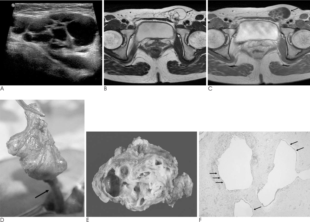
- Benign multicystic mesothelioma is a rare mesothelial lesion that forms multicystic masses in the upper abdomen, pelvis, and retroperitoneum. Most cases have a benign course. We present the ultrasound and MR findings of benign multicystic mesothelioma in the left round ligament, which caused a left inguinal hernia in a 46-year-old woman.
MeSH Terms
Figure
Reference
-
1. Safioleas MC, Constantinos K, Michael S, Konstantinos G, Constantinos S, Alkiviadis K. Benign multicystic peritoneal mesothelioma: a case report and review of the literature. World J Gastroenterol. 2006; 12:5739–5742.2. Choi YC, Choi HC, Chang TS, Kwon OJ, Kim BH. A Cystic Mesothelioma in the Right Colon: A case report. J Korean Surg Soc. 1998; 55:919–924.3. Lim SC, Jeong YK, Lee MS, Kim YS, Park HJ, Choi SJ. Benign Cystic Mesothelioma. Korean J Pathol. 1997; 31:595–597.4. Søreide JA, Søreide K, Körner H, Søiland H, Greve OJ, Gudlaugsson E. Benign peritoneal cystic mesothelioma. World J Surg. 2006; 30:560–566.5. O'Neil JD, Ros PR, Storm BL, Buck JL, Wilkinson EJ. Cystic mesothelioma of the peritoneum. Radiology. 1989; 170:333–337.6. Weiss SW, Tavassoli FA. Multicystic mesothelioma: an analysis of pathologic findings and biologic behavior in 37 cases. Am J Surg Pathol. 1988; 12:737–746.7. Ryley DA, Moorman DW, Hecht JL, Alper MM. A mesothelial cyst of the round ligament presenting as an inguinal hernia after gonadotropin stimulation for in vitro fertilization. Fertil Steril. 2004; 82:944–946.8. Kim SH, Seo IY, Cho HJ, Ku YM, Kim KH, Ahn CH, Kim JS, Yoo SJ, Lim KW, Kim JI. Hydrocele of the Canal of Nuck. J Korean Surg Soc. 2008; 74:396–398.9. Walshe JM, Gal A, Murray DR, Premkumar A, Berman D, Hassan R. Malignant mesothelioma of the inguinal canal with an unusually long survival. Am J Clin Oncol. 2008; 31:306–307.
- Full Text Links
- Actions
-
Cited
- CITED
-
- Close
- Share
- Similar articles
-
- Multicystic mesothelioma of the peritoneum: case report
- Multicystic Mesothelioma of the peritoneum: A case Report
- Multicystic benign mesothelioma of the pelvic peritoneum presenting as acute abdominal pain in a young woman
- A Case of Large Cystic Myxoid Leiomyoma in Uterine Round Ligament
- Ultrasonographic diagnosis of round ligament varicosities mimicking inguinal hernia: report of two cases with literature review


