The MDCT and MRI Findings of a Pancreatic Arteriovenous Malformation Combined with Isolated Dissection of the Superior Mesenteric Artery: A Case Report
- Affiliations
-
- 1Department of Radiology, Hanyang University Guri Hospital, Korea. ysookim@hanyang.ac.kr
- 2Department of Radiology, Naval Pohang Hospital, Korea.
- 3Department of Radiology, Hanyang University Medical College, Korea.
- KMID: 2208921
- DOI: http://doi.org/10.3348/jksr.2010.62.3.257
Abstract
- Pancreatic arteriovenous malformation and isolated spontaneous dissection of the superior mesenteric artery are both rare maladies, and now they can be easily diagnosed due to the development of such noninvasive modalities as multi-detector computed tomography and magnetic resonance imaging. We report here on the multi-detector computed tomography and magnetic resonance imaging findings of a rare case of pancreatic arteriovenous malformation combined with isolated dissection of the superior mesenteric artery.
MeSH Terms
Figure
-
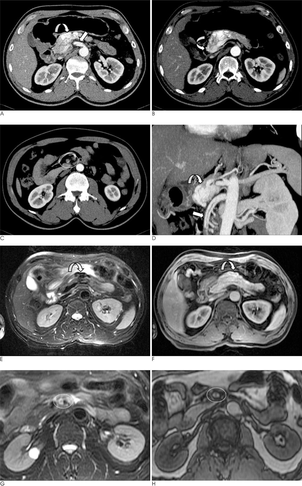
Fig. 1 The MDCT and MRI findings of a 57-year-old man with epigastric pain. A. The axial scan during the arterial phase shows early-enhancing tortuous, dilated mass of vasculature (curved arrow) in the body of the pancreas. A dissecting flap (straight arrow) is seen within the SMA, beginning just distal to its origin from the aorta. B. Early enhancement of the portal vein (curved arrow) is seen during the arterial phase. C. The false lumen (within the broken lines) is seen as the low-attenuation filling defect that is due to thrombus. D. The oblique coronal MPR image provides a clearer image of both the pancreatic AVM (curved arrow) and the SMA dissection (straight arrow). E. On the fat-suppressed T2WI, the pancreatic AVM (curved arrow) demonstrates the characteristic signal void. F. The AVM (curved arrow) shows marked enhancement on the contrast-enhanced T1WI. G. The fat-suppressed T2WI of the proximal SMA reveals a discrepancy in the signal intensities of the true and false lumens (within the broken lines). The true lumen demonstrates a signal void due to high blood flow, while the false lumen shows hyperintensity due to the slower blood flow. H. The contrast-enhanced T1WI demonstrates enhancement of both the true and false lumens of the dissected SMA (within the broken lines).
Reference
-
1. Halpern M, Turner AF, Citron BP. Hereditary hemorrhagic teleangiectasia. an angiographic study of abdominal visceral angiodysplasia associated with gastrointestinal hemorrhage. Radiology. 1968; 90:1143–1149.2. Bauersfield SR. Dissecting aneurysm of the aorta: a presentation of fifteen cases and a review of the recent literature. Ann Intern Med. 1947; 26:873–889.3. Hosogi H, Ikai I, Hatano E, Taura K, Fujii H, Uamamoto Y, et al. Pancreatic arteriovenous malformation with portal hypertension. J Hepatobiliary Pancreat Surg. 2006; 13:344–346.4. Yoon JH, Han SS, Cha SS, Lee SJ. Color Doppler ultrasonography of a pancreatic arteriovenous malformation. J Ultrasound Med. 2005; 24:113–117.5. Nishiyama R, Kawanishi Y, Mitsuhashi H, Kanai T, Ohba K, Mori T, et al. Management of pancreatic arteriovenous malformation. J Hepatobiliary Pancreat Surg. 2000; 7:438–442.6. Kurosaki M, Hattori K, Minato Y, Shiiqai T, Ohashi I, Umehara I, et al. Asymptomatic arteriovenous malformation of the pancreas: demonstration by Doppler ultrasonography and magnetic resonance imaging. Dig Dis Sci. 1993; 38:1342–1346.7. Rezende MB, Bramhall S, Hayes T, Olliff S, Buckels JA, Candinas D, et al. Pancreatic arteriovenous malformation. Dig Surg. 2003; 20:65–69.8. Leung DA, Schneider E, Kubik-Huch R, Marincek B, Pfammatter T. Acute mesenteric ischemia caused by spontaneous isolated dissection of the superior mesenteric artery: treatment by percutaneous stent placement. Eur Radiol. 2000; 10:1916–1919.9. Sparks SR, Vasquez JC, Bergan JJ, Owens EL. Failure of nonoperative management of isolated superior mesenteric artery dissection. Ann Vasc Surg. 2000; 14:105–109.10. Sagiuchi T, Asano Y, Yanaihara H, Aoki Y, Woodhams R, Hayakawa K. Three-dimensional CT in isolated dissecting aneurysm of the superior mesenteric artery: a case report. Radiat Med. 2001; 19:271–273.
- Full Text Links
-
- Actions
-
Cited
- CITED
-
- Close
- Share
-
- Similar articles
-
- Isolated spontaneous dissection of the superior mesenteric artery
- Pancreatic Arteriovenous Malformation Combined with Pseudocysts
- Isolated Spontaneous Dissection of the Proximal Superior Mesenteric Artery
- Transcatheter Arterial Embolization of Pancreatic Arteriovenous Malformation Presenting as Retroperitoneal Bleeding: A Case Report
- Spontaneous Isolated Dissection of the Celiac Artery: a Case Report
