Case Report of Imaging Analyses of the Dysplasia Epiphysealis Hemimelica (Trevor's Disease)
- Affiliations
-
- 1Department of Diagnostic Radiology, College of Medicine, Yeungnam University, Daegu, Korea. khcho@med.yu.ac.kr
- KMID: 2208819
- DOI: http://doi.org/10.3348/jksr.2013.69.2.149
Abstract
- Trevor's disease, also known as dysplasia epiphysealis hemimelica, is a rare developmental disorder presented with epiphyseal overgrowth involving one or multiple epiphyses. Here we report the radiologic findings of two cases of dysplasia epiphysealis hemimelica in a 4-year-old boy in the knee without symptom and a 10-year-old boy in the ankle with pain. The former was observed for eight years and the latter was treated with surgical resection.
MeSH Terms
Figure
-

Fig. 1 4-year-old man with Trevor's disease in the right distal femur. A. Initial oblique (A) radiograph shows multiple calcific foci in the postero-medial aspect of the epiphysis of the right distal femur (arrow). B. Sagittal sonography (B) shows epiphyseal cartilaginous overgrowth, containing multiple echogenic foci (arrow). C-E. Axial T2-weight image (C), axial T1-weight image (D) and axial enhanced T1-weight image (E) show the presence of asymmetric epiphyseal cartilaginous overgrowth (small arrows), which contain multiple ossifications, in the postero-medial aspect of the distal femur (arrows).

Fig. 2 4-year-old man with Trevor's disease in the right distal femur. Follow-up oblique radiographs after 12 months (A), 24 months (B), 54 months (C) and 8 years (D) show that calcific foci are maturated and becomes confluent with the epiphysis of the distal femur (arrows).
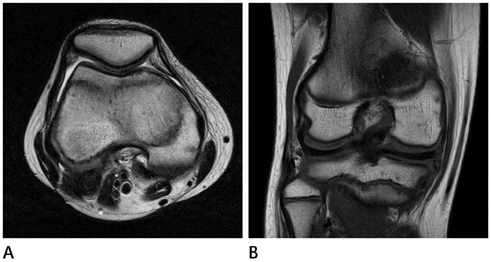
Fig. 3 4-year-old man with Trevor's disease in the right distal femur. Follow-up after 8 years, axial T2 weight image (A) and coronal T1 weight image (B) show normal bone contour and signal intensity in the distal femur with disappearance of the dysplasia epiphysealis hemimelica findings.
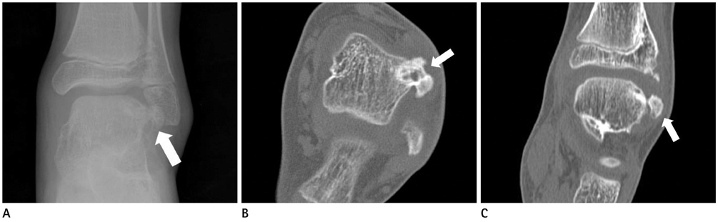
Fig. 4 10-year-old man with Trevor's disease in the left talus. Initial AP radiograph (A), unenhanced axial (B) and coronal (C) CT show irregular ossified mass, arising talus, in the antero-lateral aspect of the talus (arrows).
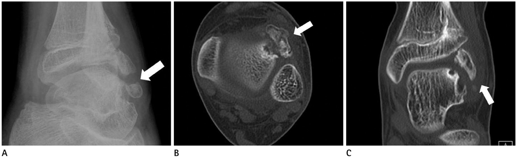
Fig. 5 10-year-old man with Trevor's disease in the left talus. Follow-up study after 2 years due to recurred ankle pain, oblique radiograph (A), unenhanced axial (B) and coronal (C) CT show recurred mass in the antero-lateral aspect of the talus (arrows).
Reference
-
1. Kuo RS, Bellemore MC, Monsell FP, Frawley K, Kozlowski K. Dysplasia epiphysealis hemimelica: clinical features and management. J Pediatr Orthop. 1998; 18:543–548.2. Shinozaki T, Ohfuchi T, Watanabe H, Aoki J, Fukuda T, Takagishi K. Dysplasia epiphysealis hemimelica of the proximal tibia showing epiphyseal osteochondroma in an adult. Clin Imaging. 1999; 23:168–171.3. Rosero VM, Kiss S, Terebessy T, Köllö K, Szöke G. Dysplasia epiphysealis hemimelica (Trevor's disease): 7 of our own cases and a review of the literature. Acta Orthop. 2007; 78:856–861.4. Tyler PA, Rajeswaran G, Saifuddin A. Imaging of dysplasia epiphysealis hemimelica (Trevor's disease). Clin Radiol. 2013; 68:415–421.5. Lang IM, Azouz EM. MRI appearances of dysplasia epiphysealis hemimelica of the knee. Skeletal Radiol. 1997; 26:226–229.6. Keret D, Spatz DK, Caro PA, Mason DE. Dysplasia epiphysealis hemimelica: diagnosis and treatment. J Pediatr Orthop. 1992; 12:365–372.
