Extraskeletal Primary Ewing's Sarcoma in the Nasal Cavity: A Case Report
- Affiliations
-
- 1Department of Radiology, Dongguk University College of Medicine, Ilsan Hospital, Goyang, Korea. ejl1048@hanmail.net
- 2Department of Otolaryngology, Dongguk University College of Medicine, Ilsan Hospital, Goyang, Korea.
- 3Department of Pathology, Dongguk University College of Medicine, Ilsan Hospital, Goyang, Korea.
- KMID: 2208812
- DOI: http://doi.org/10.3348/jksr.2013.69.2.99
Abstract
- Ewing's sarcoma presents a rare tumor of the head and neck, and even rarer in the nasal cavity and/or paranasal sinuses. We report the case of Ewing's sarcoma in the nasal cavity, as presented with nasal obstruction and epistaxis. The CT and MRI examination reveals a mass in the left nasal cavity with extension to contralateral side, ethmoidal sinus, and nasopharynx. We provide an overview of Ewing's sarcoma in the nasal cavity and discuss radiologic findings of this unusual case.
Figure
-
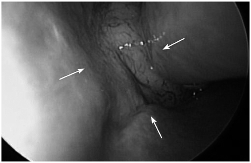
Fig. 1 Initial endoscopic features. A lobulated mass (arrows) coated with discharge was detected in the left nasal cavity expanding to contralateral side.

Fig. 2 Ewing's sarcoma in the left nasal cavity in a 42-year-old man. Postcontrast coronal CT image (A) shows an expansile and well-defined soft tissue mass (arrows) in the left nasal cavity with inhomogeneous enhancement. The mass extends to the contralateral nasal cavity, left posterior ethmoid sinus and hard palate. Coronal CT image with bone algorithm (B) demonstrates better bony destructive change. On precontrast coronal T2-weighted image with fat suppression (C), an expansile lobulated mass (arrows) in the left nasal cavity shows hyperintense signal intensity compared with the muscle. Precontrast coronal and axial T1-weighted images with fat suppression (D, E) show slightly hyperintense signal intensity. Postcontrast T1-weighted axial image with fat suppression (F) reveals heterogeneous enhancement of the mass. As seen on CT images, extension to adjacent structures with bony destructive change is also noted.
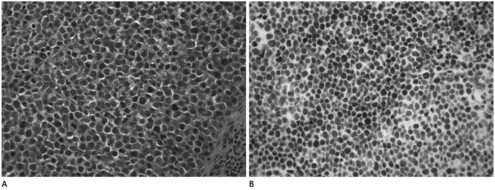
Fig. 3 Pathologic features of Ewing sarcoma/Primitive neuroectodermal tumor. A. The tumor is composed of densely distributed, uniform, small to medium sized, round cells with scanty cytoplasm. Mitotic figures are readily found (Hematoxylin & Eosin, × 400). B. The tumor cells are immunoreactive with FLI-1 protein (Immunohistochemistry, × 400). Note.-FLI-1 = Friend leukemia integration-1
Reference
-
1. Kawabata M, Yoshifuku K, Sagara Y, Kurono Y. Ewing's sarcoma/primitive neuroectodermal tumour occurring in the maxillary sinus. Rhinology. 2008; 46:75–78.2. Aferzon M, Wood WE, Powell JR. Ewing's sarcoma of the ethmoid sinus. Otolaryngol Head Neck Surg. 2003; 128:897–901.3. Harman M, Kiroğlu F, Kösem M, Unal O. Primary Ewing's sarcoma of the paranasal sinus with intracranial extension: imaging features. Dentomaxillofac Radiol. 2003; 32:343–346.4. Howarth KL, Khodaei I, Karkanevatos A, Clarke RW. A sinonasal primary Ewing's sarcoma. Int J Pediatr Otorhinolaryngol. 2004; 68:221–224.5. Javery O, Krajewski K, O'Regan K, Kis B, Giardino A, Jagannathan J, et al. A to Z of extraskeletal Ewing sarcoma family of tumors in adults: imaging features of primary disease, metastatic patterns, and treatment responses. AJR Am J Roentgenol. 2011; 197:W1015–W1022.6. Csokonai LV, Liktor B, Arató G, Helffrich F. Ewing's sarcoma in the nasal cavity. Otolaryngol Head Neck Surg. 2001; 125:665–667.7. Yeshvanth SK, Ninan K, Bhandary SK, Lakshinarayana KP, Shetty JK, Makannavar JH. Rare case of extraskeletal Ewings sarcoma of the sinonasal tract. J Cancer Res Ther. 2012; 8:142–144.8. Coskun BU, Cinar U, Savk H, Basak T, Dadas B. Isolated maxillary sinus Ewing's sarcoma. Rhinology. 2005; 43:225–228.9. Gupta S, Gupta OP, Mehrotra S, Mehrotra D. Ewing sarcoma of the maxilla: a rare presentation. Quintessence Int. 2009; 40:135–140.10. Iezzoni JC, Mills SE. "Undifferentiated" small round cell tumors of the sinonasal tract: differential diagnosis update. Am J Clin Pathol. 2005; 124:S110–S121.
