J Korean Radiol Soc.
1998 Mar;38(3):503-506. 10.3348/jkrs.1998.38.3.503.
Differential Diagnosis of the Pancreatic Diseases: Significance of Perivascular Changes at Celiac trunk andSuperior Mesenteric Artery on CT
- Affiliations
-
- 1Department of Diagnostic Radiology, Yonsei University College of Medicine.
- 2Department of Diagnostic Radiology, Sanggye Paik Hospital, Inje University College of Medicine.
- KMID: 2201409
- DOI: http://doi.org/10.3348/jkrs.1998.38.3.503
Abstract
-
PURPOSE: To classify perivascular change in the celiac trunk and SMA occurring in pancreatic disease and toevaluate its significance in differential diagnosis.
MATERIALS AND METHODS
In 73 patients with pancreaticdisease (42, acute pancreatitis; 14, chronic pancreatitis; 17, panreatic cancer) abdominal CT findings wereretrospectively reviewed. We defined " infiltration" as linear or irregular density and "thickening" as presenceof a soft tissue mantle surrounding the vessel, and statistically evaluated the usefulness of these factors forthe differential diagnosis of pancreatic diseases.
RESULTS
In 13/42 cases of acute pancreatitis (31%), 4/14 ofchronic pancreatitis (28.6%), and 6/17 of pancreatic cancer (35.3%), periceliac infiltration was observed; thefrequencies were not statistically significant (p=0.916). Peri-SMA infiltration was demonstrated in 9/42 of acutepancreatitis (21.4%), 4/14 of chronic pancreatitis (28.6%), and 5/17 of pancreatic cancer (29.4%); again, thesefrequencies were not statistically significant (p=0.758). Thickening of the celiac trunk and SMA was observed onlyin pancreatic cancer, in 3/17 (17.6%) and 7/17(41.2%) cases, respectively, with statistical significance (p<0.05).
CONCLUSION
Thickening of the celiac trunk and SMA is a valuable finding in the differential diagnosis ofpancreatic inflammatory disease and pancreatic cancer. When applied to the differential diagnosis of pancreaticdisease, perivaseular change should be classified as either infiltration or thickening.
MeSH Terms
Figure
-
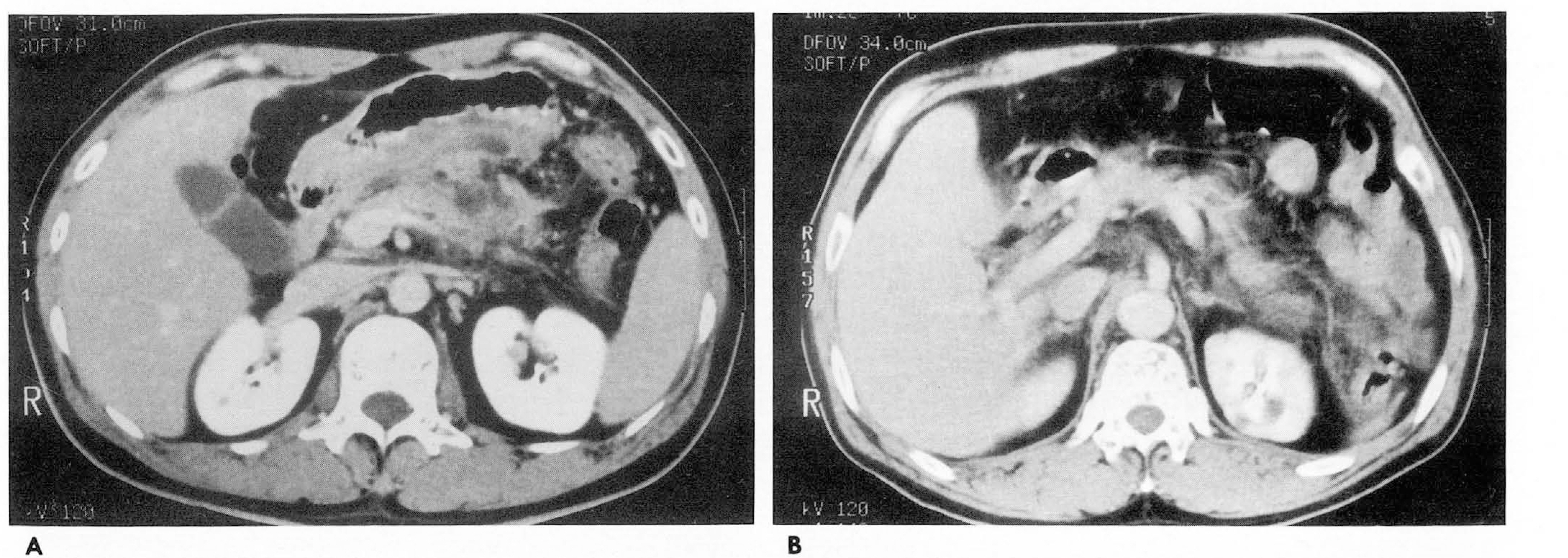
Fig. 1. A. Reticular infiltration of perivascular fat is demonstrated at SMA root in chronic pancreatitis 8. Perivascular fat infiltration is shown at celiac root in acute pancreatitis.
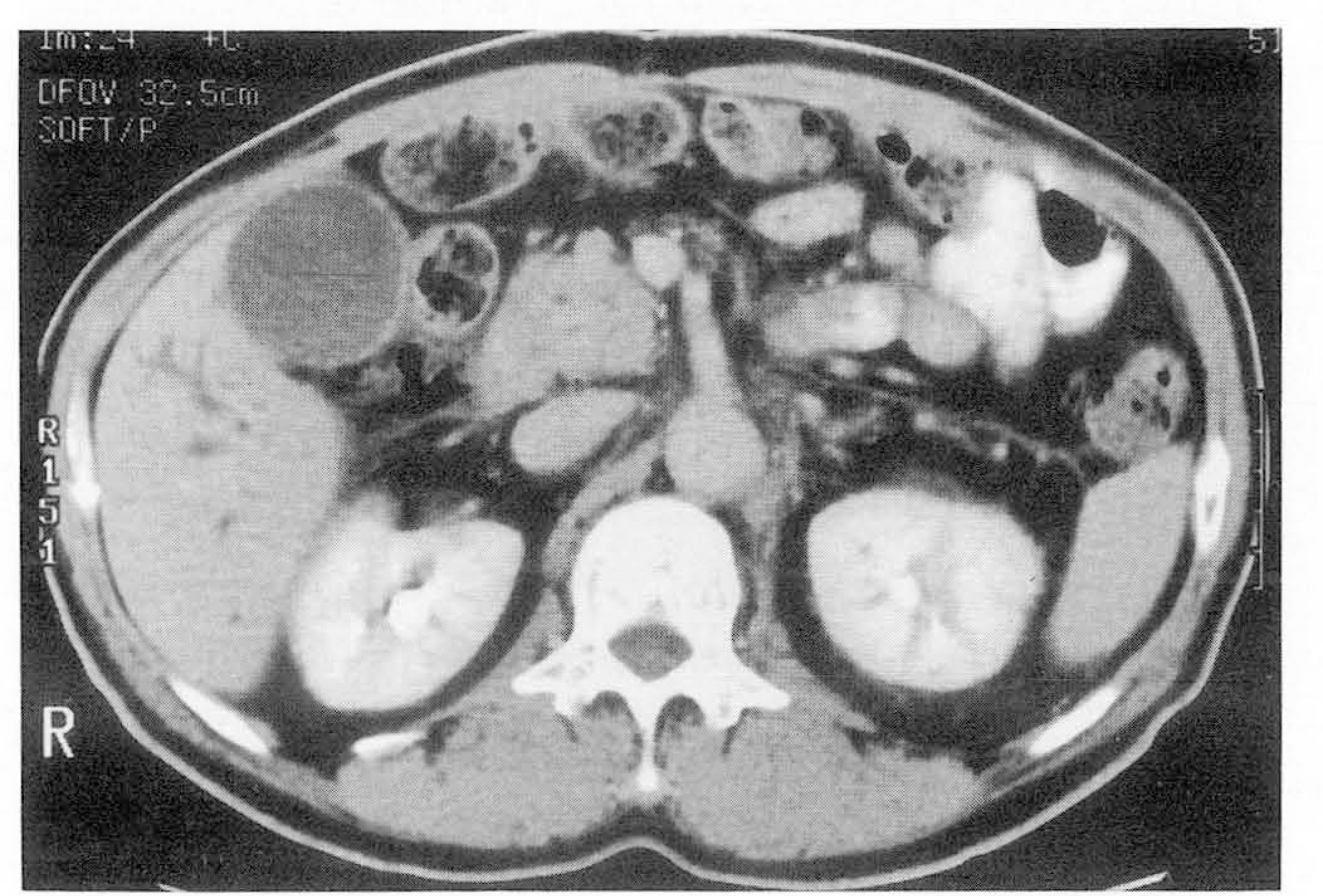
Fig. 2. Sixty-three year old male patient with pancreatic carcinoma å irregular infiltration is noted at SMA root area.
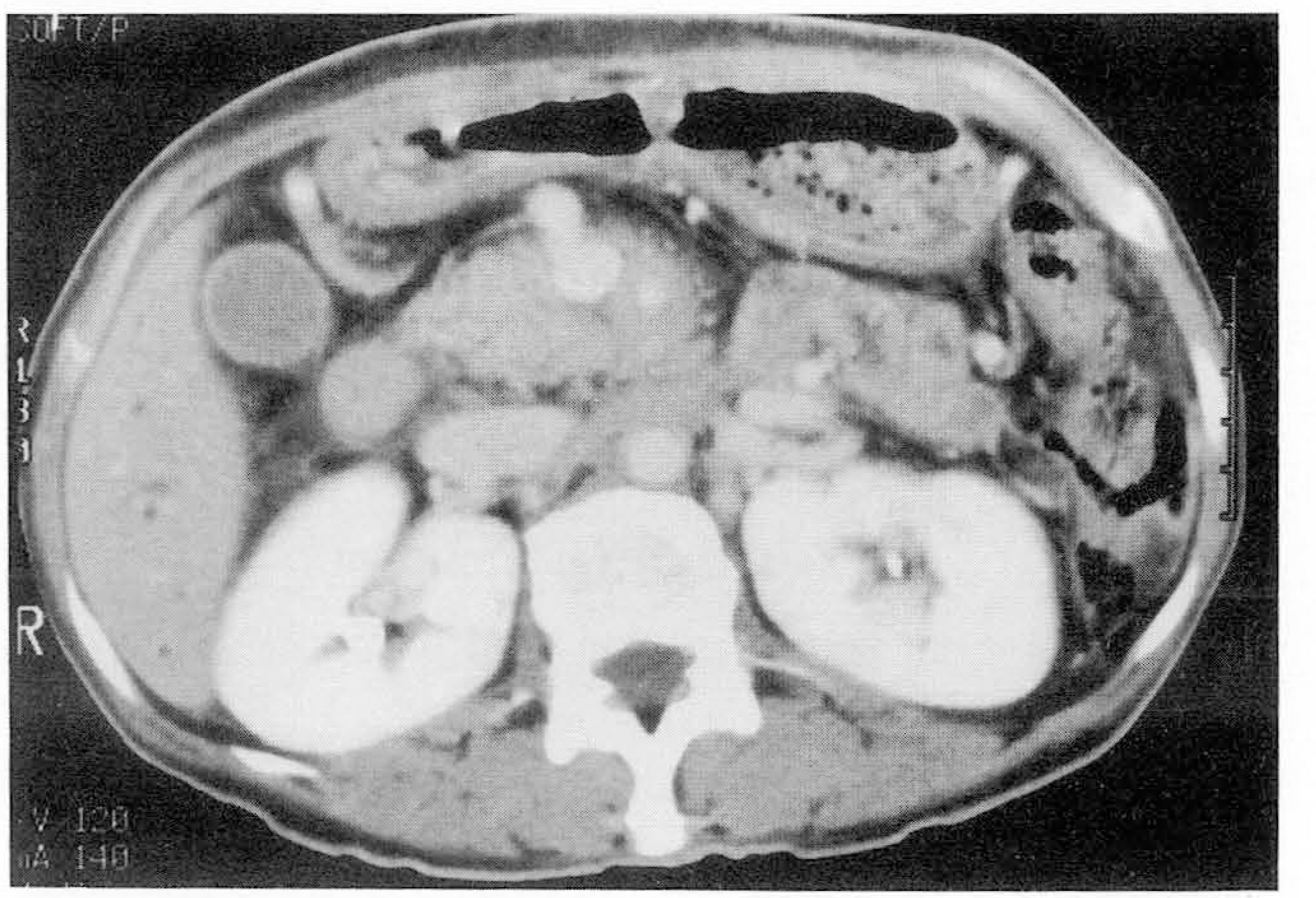
Fig. 3. Fifty-three year old female patient with pancreatic carcinoma å soft tissue material is demonstrated around SMA (thickened vessel).
Reference
-
1.Megibow A J., Bostniak MA., Ambos MA., Beranhaum ER. Thickening of the celiac axis and/or superior mesenteric artery: A sign of pancreatic carcinoma on CT. Radiology. 1981. 14:449–453.2.Mitchell DG., Hill MC., Cooper R, et al. The superior mesenteric artery fat plane: Is obliteration pathognomonic of pancreatic carcinoma? J Comput Assist Tomogr. 1987. 11(3):247–253.
Article3.Baker ME., Cohan RH., Nadel SN., Leder RA., Dunnick NR. Obliteration of the Fat Surrounding the Celiac Axis and Superior Mesenteric Artery Is Not a Specific CT Fundings of Carcinoma of the Pancreas. AJR. 1990. 155:991–994.4.Luetmer PH., Stephens DH., Fischer AP. Obliteration of periarterial retropancreatic fat on CT in pancreatitis: an exception to the rule. AJR. 1989. 153:63–64.
Article5.Schulte SJ., Baron RL., Freeny PC., Patten RM., Gorell HA., Maclin ML. Root of the Superior Mesenteric Artery in Pancreatitis and Pancreatic Carcinoma: Evaluation with CT. Radiology. 1991. 180:659–662.
Article6.Baker ME. Pancreatic Adenocarcinoma: Are There Pathognomonic Changes in the Fat Surrounding the Superior Mesenteric Artery? Radiology. 1991. 180:613–614.
Article7.Gortenuti G., Cavallini G,Vantini I., Angelini G. Angiography in chronic pancreatitis and pancreatic cancer. Am J Gastroenterol. 1978. 70:620–626.8.Megibow AJ. Pancreatic Adenocarcinoma: Designing the Examination to Evaluate the Clinical Questions. Radiology. 1992. 183:297–303.
Article9.Cubilla AL., Fortner J., Fitzgerald Ρ J. Lymph node involvement in carcinoma of the head of the pancreas area. Cancer. 1978. 41:880–887.
Article10.Balthazar EJ. CT diagnosis and staging of acute pancreatitis. Radiol Clin North Am. 1989. 27:19–37.
- Full Text Links
-
- Actions
-
Cited
- CITED
-
- Close
- Share
-
- Similar articles
-
- Variations in the branching pattern of the celiac trunk and its clinical significance
- Isolated Bypass to the Superior Mesenteric Artery for Chronic Mesenteric Ischemia
- A Rare Case in the Pattern of the Origin of the Celiac Artery
- Endovascular Treatment of Chronic Mesenteric Ischemia by Crossing of Two Stents in a Patient with Celiacomesenteric Trunk
- A rare variant angioarchitecture of upper abdomen
