Qualitative Analysis of Barium Particles Coated in Small Intestinal Mucosa of Rabbit by Using Scanning ElectronMicroscopy
- Affiliations
-
- 1Department of Diagnostic Radiology, Asan Medical Center, University of Ulsan College of Medicine.
- KMID: 2201406
- DOI: http://doi.org/10.3348/jkrs.1998.38.3.485
Abstract
-
PURPOSE: To qualitatively analyse barium coating status in the intestinal mucosa, we used scanning electronmicroscopy to observe barium particles coated in the small intestinal mucosa of rabbit, and we attempted to assessthe relationship between electron microscopic findings and radiographic densities.
MATERIALS AND METHODS
Sixdifferent combinations of barium and methylcellulose suspensions were infused into the resected small intestinesof 15 rabbits. Barium powders were mixed with water to make 40% and 70% w/v barium solutions, and also mixed with0.5% methylcellulose solution to make 40% and 70% w/v barium-methylcellulose mixtures. 0.5% methylcellulosesolutions were used as a double contrast agent. After the infusion of barium suspensions, a mammography unit wasused to obtain radiographs of the small intestine, and their optical densities were measured by a densitometer.Thereafter, photographs of barium-coated small intestinal mucosa were obtained using a scanning electronmicroscope (x8,000), and the number of barium particles in the unit area were measured. To compare therelationship between the electron microscopic findings and optical densities, statistical analysis using Spearmancorrelation was performed.
RESULTS
With a Spearman coefficient of-0.544, correlation between the number of smallbarium particles of less than 1 micrometer and optical densities was statistically significant(p<0.05).
CONCLUSION
Thisstudy shows that by using scanning electron microscopy, barium particles coated on the small intestinal mucosa canbe qualitatively analysed. It also shows that the number of small barium particles measured by scanning electronmicroscopy is related to optical densities.
Keyword
MeSH Terms
Figure
-
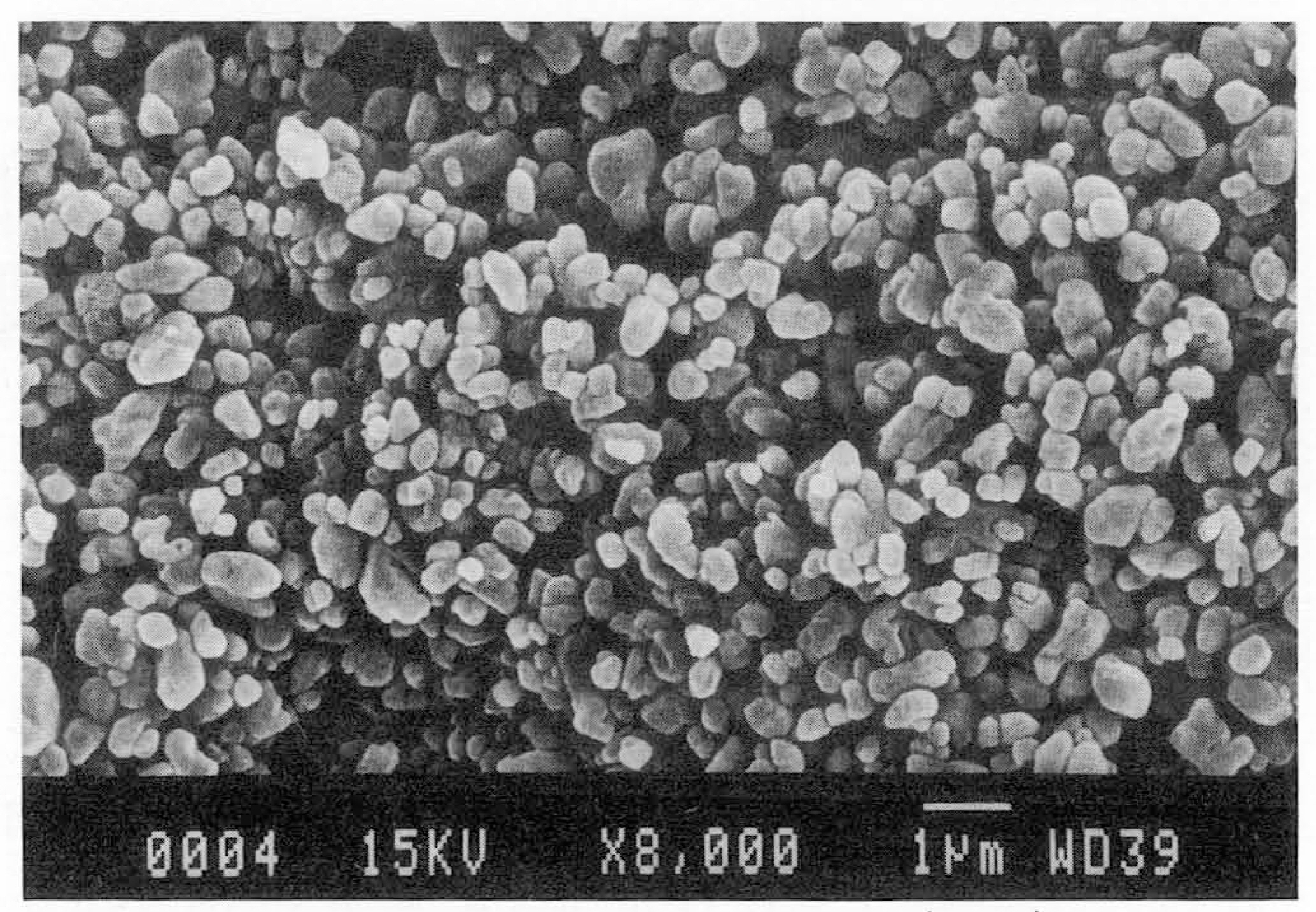
Fig. 1. Scanning electron microscopy (SEM) of barium suspension(X 8,000). Barium particles show variable size and shape, but most of them are less than 1 //m. Aggregations are rarely seen.

Fig. 2. A. SEM of group 1(X 8,000). Most of barium particles are large particles, and small barium particles are rarely seen. B. Radiography of group 1. A radiograph using mammographic technique shows aggregation of barium particles with reticular appearance. C. SEM of group 2(X 8,000). Compared to Fig. 2A, the number of small barium particles are increased. D. Radiography of group 2. Similar reticular patterns are also seen as in group 1. E. SEM of group 3(X 8,000). Small barium particles are densely coated. Particles vary in size and shape, and many particles are aggregated. F. Radiography of group 3. Due to a large amount of barium particles, mucosal details are not seen at all on this very radiopaque radiograph.
Reference
-
1.Maglinte DDT., Lappas JC., Kelvin FM., Rex D., Chernish SM. Small bowel radiography: how, when, and why? Radiology. 1987. 163:297–305.
Article2.Herlinger H. Guide to imaging of the small bowel. Gastroenterol Clin North Am. 1995. 24:309–329.
Article3.Klein HM., Gunther RW. Double contrast small bowel follow-through with an acid-resistant effervescent agent. Invest Radiol. 1993. 7:581–585.
Article4.Fitch D. The small bowel see-through: an improved method of radiographic small-bowel visualization. Can J Med Radiat Technol. 1995. 26:167–171.5.Skucas J. Contrast media. In Gore RM, Levine MS, Laufer I, eds. Textbook of Gastrointestinal Radiology. Philadelphia: Saunders. 1994. 17–30.6.Salomonᄋwitz E., Frick MP., Cragg AH., Lund G. The adhesiometer: a simple device to measure adherence of barium sulfate to intestinal mucosa. AJR. 1984. 142:721–723.7.서태석, 이동호, 고영태, 임주원, 한태일, 김형중. 이중조영바륨관장검사에서바륨현탁액의대장점막도포정도: 증류수와생리식염수제재의비교. 대한방사선의학희지. 1997. 36:1029–1032.8.Antes G., Lissner J. Double-contrast small-bowel examination with barium and methylcellulose. Radiology. 1983. 148:37–40.
Article9.Ott DJ., Chen YM., Gelfand DW., Swearingen FV., Munitz HA. Detailed per-oral small bowel examination vs. enteroclysis. Radiology. 1985. 155:29–34.10.Phillips JK., Scott RL., Nicell DT. The magic tilt method of tubeless enteroclysis: a modification to the gas-enhanced barium small-bowel examination. AJR. 1996. 166:358–359.
Article11.Herlinger H. Small bowel. In Laufer I, Levine MS, eds. Double contrast gastrointestinal radiology. 2nd ed.Philadelphia: Saunders;1992. p. 363–422.12.Desaga JF. Visualization of the mucosal villi on double-contrast barium studies of the small intestine by using a high molecular fraction of guaran. Gastrointest Radiol. 1989. 14:25–30.
Article13.Gelfand DW., Ott DJ. Barium sulfate suspension: an evaluation of a available products. AJR. 1982. 138:935–941.14.Schwarz SE., Fischer HW., House AJF. Studies in adherence of contrast media to mucosal surfaces. Radiology. 1974. 112:727–731.
Article
