Radiologic Findings of Acute Spontaneous Subdural Hematomas
- Affiliations
-
- 1Department of Radiology, Chonan Hospital Soonchunhyang University.
- 2Department of Neurosurgery, Chonan Hospital Soonchunhyang University.
- KMID: 2201391
- DOI: http://doi.org/10.3348/jkrs.1998.38.3.391
Abstract
-
PURPOSE: To evaluate the characteristic CT and cerebral angiographic findings in patients with acutespontaneous subdural hematomas and correlate these imaging findings with causes of bleeding and clinical outcome.
MATERIALS AND METHODS
Twenty-one patients with nontraumatic acute spontaneous subdural hematoma presentingduring the last five years underwent CT scanning and cerebral angiography was performed in twelve. To determinethe cause of bleeding, CT and angiographic findings were retrospectively analysed. Clinical history, laboratoryand operative findings, and final clinical outcome were reviewed.
RESULTS
The 21 cases of acute spontaneoussubdural hematomas were caused by cerebral vascular abnormalities(n=10), infantile hemorrhagic disease(n=5), orwere of unknown origin(n=6). All ten cases of cerebral vascular abnormality were confirmed angiographically; sixwere aneurysms, three were arteriovenous malformations, and one was moyamoya disease. On CT, subarachnoidhemorrhage was seen to be associated with aneurysms, intracerebral hemorrhage with arteriovenous malformations,and intraventricular hemorrhage with moyamoya disease. All five patients with hemorrhagic disease were infantsaged 1-17 months ; characteristic diffuse distribution of subdural hematoma in both temporoparietal-occipitalregions is typical. The average overall mortality rate was 52.4%(11/21). In patients with cerebral vascularabnormalities, mortality was as low as 20%(2/10), but in hemorrhagic disease was high (60%). In cases of unknownorigin it was 100%.
CONCLUSION
Acute spontaneous subdural hematoma is a rare condition, and the mortality rateis high. In patients with acute spontaneous subdural hematoma, as seen on CT, associated subarachnoid orintracerebral hemorrhage is strongly indicative of intracerebral vascular abnormalities such as aneurysm andarteriovenous malformation, and cerebral angiography is necessary. To ensure proper treatment and thus morkedlyreduce mortality, the causes of bleeding should be prompty determined by means of cerebral angiography.
MeSH Terms
Figure
-

Fig. 1. 56 year-old female (Case No. 2) with the aneurysm. A. Noncontrast CT scan shows diffuse subarachnoid hemorrhage and thin subdural hematoma in both frontal repions. B. Left internal carotid angiogram shows small round aneurysm (arrowheads) on the bifurcation of left miadle cerebral artery.
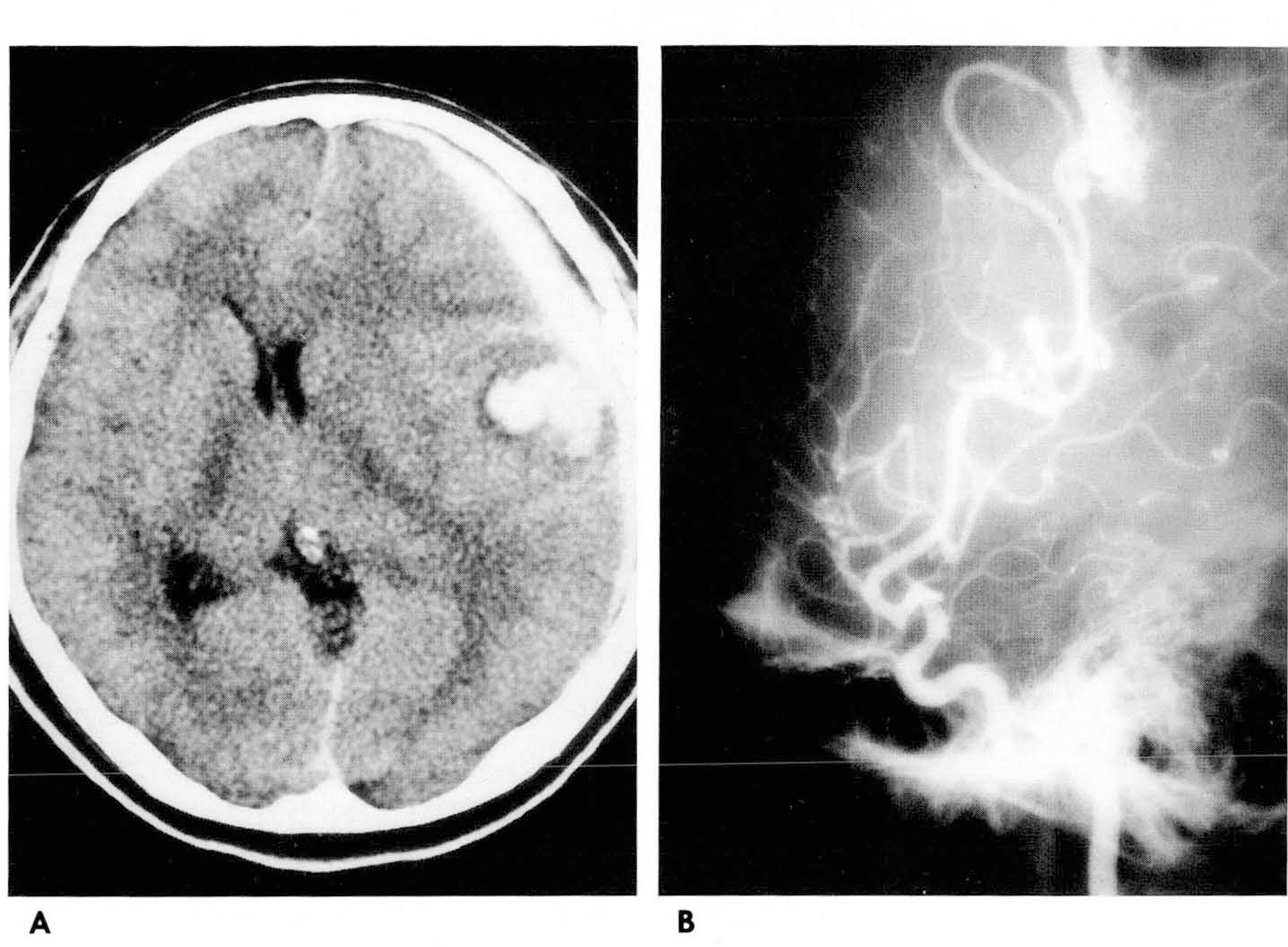
Fig. 2. 24 year-old female (Case No. 8) with the arteriovenous malformation. A. Noncontrast CT scan shows thick subdural hematoma on the left frontotemporal region and some intracerebral hematoma in adjacent cortical region. Mild midline shifting is seen to the right side. B. Left internal carotid angiogram shows the nidus of small arteriovenous malformation feeding from precentral branch of the left middle cerebral artery.
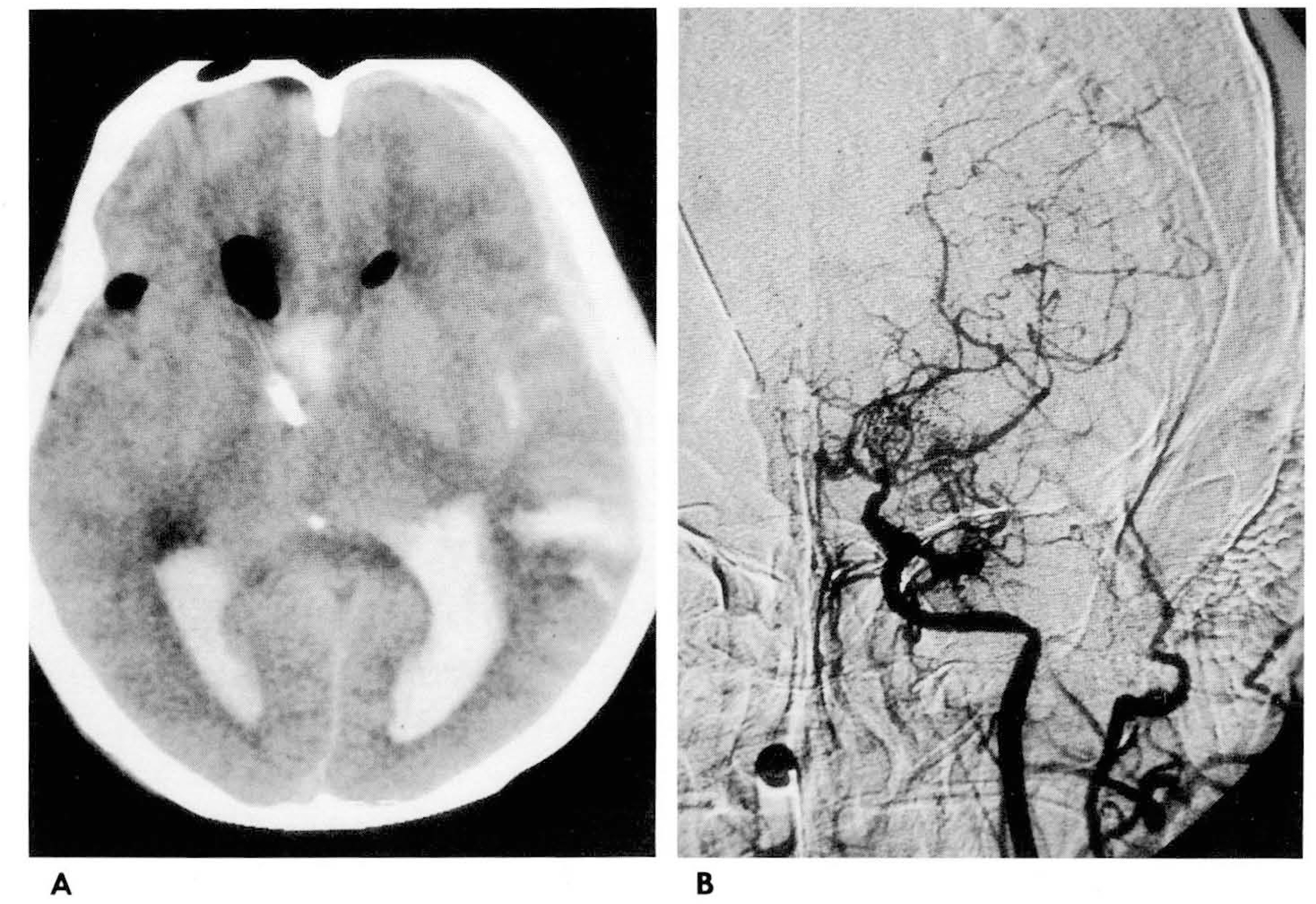
Fig. 3. 59 year-old female (Case No. 10) with moyamoya disease. A. Noncontrast CT scan shows thin acute subdural hematoma on left frontotemporal region with intraventricular hemorrhage and subarac- hmoid hemorrhage. Ventricular shunt catheter was inserted. B. Left internal carotid angiogram shows occlusion of distal internal cerebral artery with some moyamoy- a vessels. Right cartoid angiogram (not shown) shows similar findings as left.
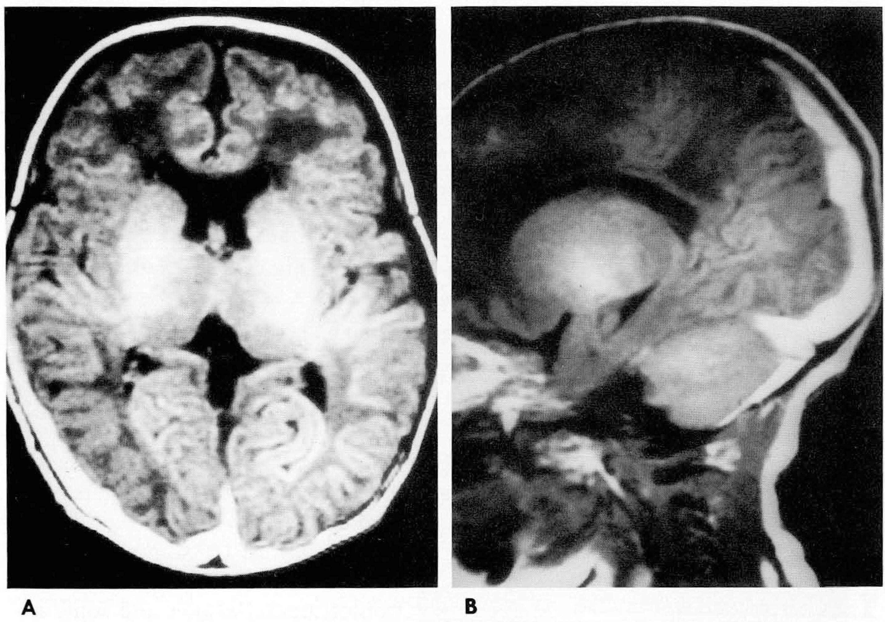
Fig. 4. 1.7 month-old female (Case No. 15) with hemorrhagic disease. Axial (A) and sagittal (B) Tl- weighted spin echo images show diffuse subdural hematoma in both occipitotemporoparietal region.
Reference
-
1.O'Brien PK., Norris JW., Tator CH. Acute subdural hematoma of arterial origin. J Neurosurg. 1974. 41:435–349.2.Byun HS., Patel PP. Spontaneous subdural hematoma of arterial origin: report of two cases. Neurosurgery. 1979. 5:611–613.3.Rengachary SS., Szymansky DC. Subdural hematomas of arterial origin. Neurosurgery. 1981. 8:166–172.
Article4.Yanai Y., Kohono N., Mitsui T. Acute spontaneous subdural hematoma of arterial origin. Surg Neurol. 1985. 23:417–420.
Article5.Borzone M., Altomonte M., Baldini M., Rivano C. Pure subdural hematomas of arterial origin. Acta NeurochirfWien). 1993. 121:109–112.6.Arienta C., Ceretti L., Caroli M., Villani R. Acute spontaneous subdural hematomas. J Neurosurg Sci. 1986. 30:197–204.7.Avis SP. Nontraumatic acute subdural hematoma. Am J Forensic Med Pathol. 1993. 14:130–134.
Article8.Kamiya K., Inagawa T., Yamamoto M., Monden S. Subdural hematoma due to ruptured intracranial aneurysm. Neurol Med Chir (Tokyo). 1991. 31:82–86.
Article9.Barton E., Tudor J. Subdural hematoma in association with intracranial aneurysm. Neuroradiology. 1982. 23:157–160.10.Pasqualin A., Bazzan A., Cavazzini P., Scienza R., Licata C., Dapian R. Intracranial hematomas following aneurysmal rupture: experience with 309 cases. Surg Neurol. 1986. 25:6–17.
Article11.Reynolds AF., Shaw C. Bleeding patterns from ruptured intracranial aneurysms: an autopsy series of 205 patients. Surg Neurol. 1981. 15:232–235.
Article12.Ragland RL., Gelber ND., Wilkinson HA., Knorr JR., Tran AA. Anterior communicating artery aneurysm rupture: an unusual cause of acute subdural hemorrhage. Surg Neurol. 1994. 40:400–402.
Article13.Kondziolka D., Bernstein M., Brugge K., Schutz H. Acute subdural hematoma from ruptured posterior communicating artery aneurysm. Neurosurgery. 1988. 22:151–154.
Article14.Wtanabe K., Wakai S., Okuhata S., Nagai M. Ruptured distal anterior cerebral artery aneurysms presenting as acute subdural hematoma: report of three cases. Neurol Med Chir (Tokyo). 1991. 31:514–517.15.Oikawa A., Aoki N., Sakai T. Arteriovenous malformation presenting as acute subdural hematoma. Neurol Res. 1993. 15:353–355.16.Kawakami K., Takahashi S., Sonoba M., Koshuk K., Hirota S., Kusunoso M. Subacute subdural hematoma associated with moyamoya phenomenon: a case report. No Shinkei Geka. 1988. 16:205–209.17.Oppenheim JS., Gennuso R., Sacher M., Hollis P. Acute atraumatic subdural hematoma associated with Moyamoya disease in an African-American. Neurosurgery. 1991. 28:616–618.
Article18.Tokoro K., Nakajima F., Yamataki A. Acute spontaneous subdural hematoma of arterial origin. Surg Neufol. 1988. 29:159–163.
Article19.Chaou WT., Chou ML., Eitzman DV. Intracranial hemorrhage and vitamin Κ deficiency in early infancy. J Pediatr. 1984. 105:880–884.20.최상호, 최재유, 김지성, 강임주. 두개내출혈을동반한영아기출혈성질환6예. 대한소아과학회지. 1988. 31:106–112.
- Full Text Links
-
- Actions
-
Cited
- CITED
-
- Close
- Share
-
- Similar articles
-
- Spontaneous Concomitant Intracranial and Spinal Subdural Hematomas in Association with Anticoagulation Therapy
- Inhospital Spontaneous Acute Subdural Hematoma (SADH) Patient with Antiplatelet Therapy due to Acute Cerebral Ischemia: A Case Report
- Computed tomography (CT) of the spontaneous resolution of traumatic epidural and subdural hematoma
- Acute Subdural Hematomas Review of 100 Cases
- Evolution of Chronic Subdural Hematoma based on Brain CT findings and Appropriate Treatment Methods
