J Lung Cancer.
2009 Dec;8(2):111-113. 10.6058/jlc.2009.8.2.111.
Primary Lung Adenocarcinoma Metastasis to the Vagina: A Case Report
- Affiliations
-
- 1Department of Pathology, Gachon University Gil Medical Center, Incheon, Korea. syha@gilhospital.com
- 2Department of Internal Medicine, Gachon University Gil Medical Center, Incheon, Korea.
- 3Department of Obstetrics and Gynecology, Gachon University Gil Medical Center, Incheon, Korea.
- 4Department of Pathology, Sungkyunkwan University Samsung Medical Center, Seoul, Korea.
- KMID: 2199971
- DOI: http://doi.org/10.6058/jlc.2009.8.2.111
Abstract
- Lung cancer is a malignant tumor that is often fatal. Vaginal metastasis of pulmonary adenocarcinoma is very rare. To the best of our knowledge, this is the second such report worldwide and the first one from Korea. A 67-year-old woman presented with cough, excessive sputum and dyspnea that she had sufferd with for the past one year and she had a palpable lesion in the vagina. Chest CT showed diffuse bronchial wall thickening involving the left main bronchus, the left upper lobar bronchus and the lingular divisional bronchus of the left upper lobe. There were multiple, various sized nodules in both lungs, of which the largest one measured about 1.0 cm in diameter. Both lung and vaginal biopsies were performed and the masses were diagnosed as adenocarcinoma. Immunohistochemically, the tumor cells were positive for cytokeratin 7 and TTF-1, but they were negative for cytokeratin 20. We present this case of primary lung adenocarcinoma metastasis to the vagina.
Keyword
MeSH Terms
Figure
-
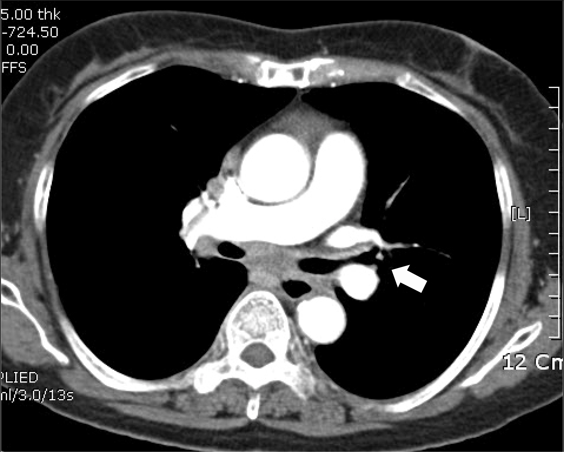
Fig. 1. Chest CT showed the bronchial wall thickening involving the left main bronchus and the left upper lobar bronchus (arrow).
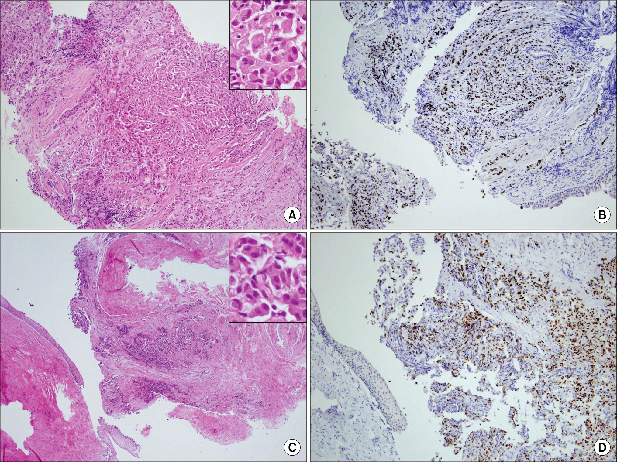
Fig. 2. The histological findings for the lung mass: bronchoscopic biopsy showed infiltration of atypical ovoid cells with round nuclei and plump cytoplasm (inlet) (A, H&E stain, ×40). The tumor cells were positive for TTF-1 (B, TTF-1, ×200). The histological findings for the tumor cells in the vagina: microscopically, the same atypical cells of the lung (inlet) tumor had infiltrated into the vaginal stroma (C, H&E stain, ×40). These cells revealed a positive reaction for TTF-1 (D, TTF-1, ×200).
Reference
-
References
1. Rosen ST, Aisner J, Makuch RW, et al. Carcinomatous leptomeningitis in small cell lung cancer: a clinicopathologic review of the National Cancer Institute experience. Medicine (Baltimore). 1982; 61:45–53.2. National Cancer Information Center. NCIC Cancer Information Service [Internet]. Goyang (Korea): National Cancer Information Center;2008. Nov 13. Available from:. http://www.cancer.go.kr/cms/statics/incidence.3. Kumar V, Abbas AK, Fausto N, Robbins SL, Cotran RS. Robbins and Cotran pathologic basis of disease. 7th ed.Philadelphia: Elsevier Saunders;2005.4. Jahnke A, Domke R, Makovitzky J, Nizze H, Briese V. Vaginal metastasis of lung cancer: a case report. Anticancer Res. 2005; 25:1645–1648.5. Mazur MT, Hsueh S, Gersell DJ. Metastases to the female genital tract. Analysis of 325 cases. Cancer. 1984; 53:1978–1984.
Article6. Fader AN, Brainard JA, Rose PG. Symptomatic vaginal bleeding in a postmenopausal woman: a case report of pancreatic adenocarcinoma metastasizing exclusively to the vagina. Am J Obstet Gynecol. 2007; 197:e8–9.
Article7. Saad RS, Liu YL, Han H, Landreneau RJ, Silverman JF. Prognostic significance of thyroid transcription factor-1 expression in both early-stage conventional adenocarcinoma and bronchioloalveolar carcinoma of the lung. Hum Pathol. 2004; 35:3–7.
Article8. Geldof AA. Models for cancer skeletal metastasis: a reappraisal of Batson's plexus. Anticancer Res. 1997; 17:1535–1539.
- Full Text Links
-
- Actions
-
Cited
- CITED
-
- Close
- Share
-
- Similar articles
-
- Ovarian and Pituitary Metastasis from Adenocarcinoma of the Lung: A Case Report
- Gastric Metastasis of Primary Lung Adenocarcinoma Mistaken for Primary Gastric Cancer
- A Case of Eyeball Metastasis of Lung Adenocarcinoma Confirmed by Enucleation
- Unusual Left Ventricular Endocardial Metastasis from Primary Lung Cancer
- Two Cases of Uveal Metastasis of Lung Cancer with Systemic Metastasis
