J Korean Soc Surg Hand.
2014 Sep;19(3):145-149. 10.12790/jkssh.2014.19.3.145.
Melorheostosis of the Trapezium
- Affiliations
-
- 1Department of Orthopedic Surgery, The Catholic University of Korea, Seoul, Korea.
- 2Department of Hospital Pathology, The Catholic University of Korea, Seoul, Korea. jylos1@gmail.com
- KMID: 2194136
- DOI: http://doi.org/10.12790/jkssh.2014.19.3.145
Abstract
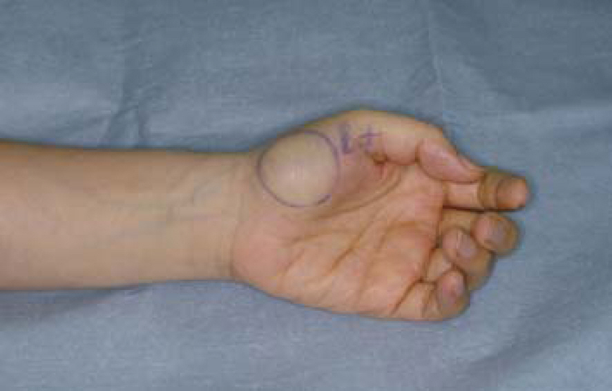
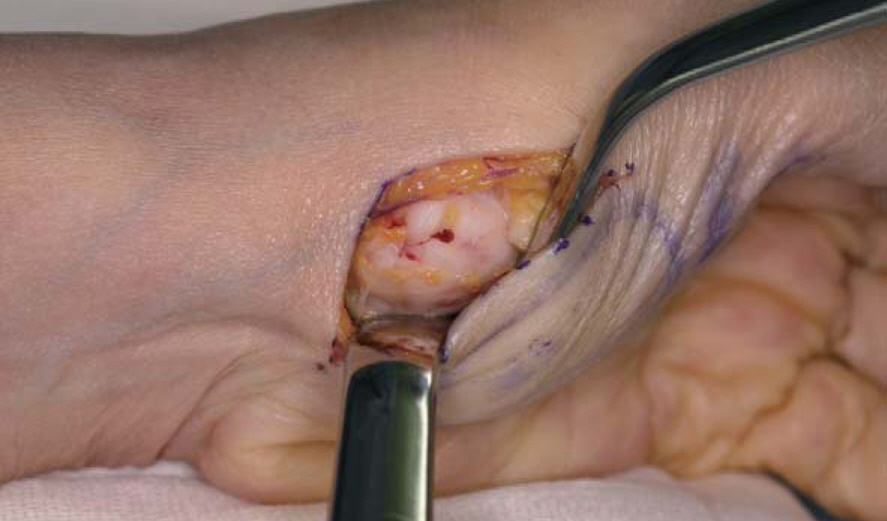
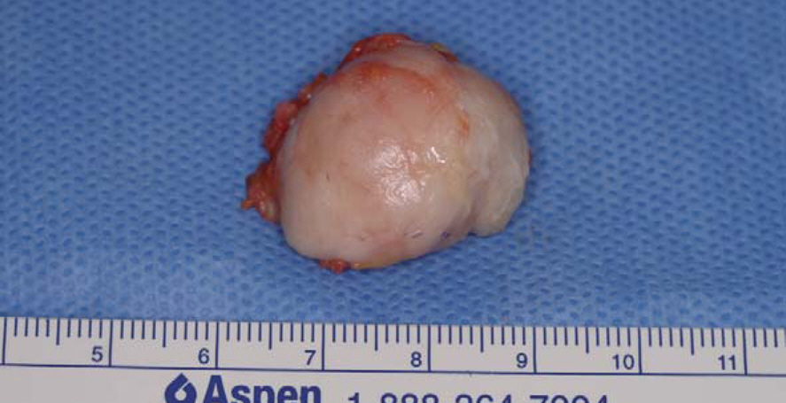
- We report a 56-year-old female with symptomatic protrusion of the bony lesion in the trapezium. Excision and biopsy of the bony lesion revealed thickened and sclerotic bony trabecula with adjacent zone of fibrocartilage, which is comparable with melorheostosis. This lesion with unique radiologic and histologic findings may be important to differentiate with other bony lesions such as myositis ossifications and osteosarcoma.
Keyword
Figure
Reference
-
1. Greenspan A, Azouz EM. Bone dysplasia series. Melorheostosis: review and update. Can Assoc Radiol J. 1999; 50:324–30.2. Jain VK, Arya RK, Bharadwaj M, Kumar S. Melorheostosis: clinicopathological features, diagnosis, and management. Orthopedics. 2009; 32:512.
Article3. Happle R. Melorheostosis may originate as a type 2 segmental manifestation of osteopoikilosis. Am J Med Genet A. 2004; 125A:221–3.
Article4. Freyschmidt J. Melorheostosis: a review of 23 cases. Eur Radiol. 2001; 11:474–9.
Article5. Rhee SK, Song SW, Lee WS, Hong SH. Melorheostosis in hand: 2 cases of report. J Korean Soc Surg Hand. 2001; 6:205–8.6. Jung ST, Jung SN, Lee KB. Melorheostosis of the foot: a case report. J Korean Orthop Assoc. 2000; 35:177–80.7. Younge D, Drummond D, Herring J, Cruess RL. Melorheostosis in children. Clinical features and natural history. J Bone Joint Surg Br. 1979; 61-B:415–l8.
Article8. Judkiewicz AM, Murphey MD, Resnik CS, Newberg AH, Temple HT, Smith WS. Advanced imaging of melorheostosis with emphasis on MRI. Skeletal Radiol. 2001; 30:447–53.
Article9. Hoshi K, Amizuka N, Kurokawa T, Nakamura K, Shiro R, Ozawa H. Histopathological characterization of melorheostosis. Orthopedics. 2001; 24:273–7.
Article10. Abdullah S, Mat Nor NF, Mohamed Haflah NH. Melorheostosis of the hand affecting the c6 sclerotome and presenting with carpal tunnel syndrome. Singapore Med J. 2014; 55:e54–6.
Article


