Circulating Vascular Progenitor Cells in Moyamoya Disease
- Affiliations
-
- 1Department of Neurosurgery, Seoul National University Hospital, Seoul National University College of Medicine, Seoul, Korea. nsthomas@snu.ac.kr
- 2Division of Pediatric Neurosurgery, Seoul National University Children's Hospital, Seoul National University College of Medicine, Seoul, Korea.
- KMID: 2191249
- DOI: http://doi.org/10.3340/jkns.2015.57.6.428
Abstract
- Various approaches have been attempted in translational moyamoya disease research. One promising material for modeling and treating this disease is vascular progenitor cells, which can be acquired and expanded from patient peripheral blood. These cells may provide a novel experimental model and enable us to obtain insights regarding moyamoya disease pathogenesis. We briefly present the recent accomplishments in regard to the studies of vascular progenitor cells in moyamoya disease.
Figure
-
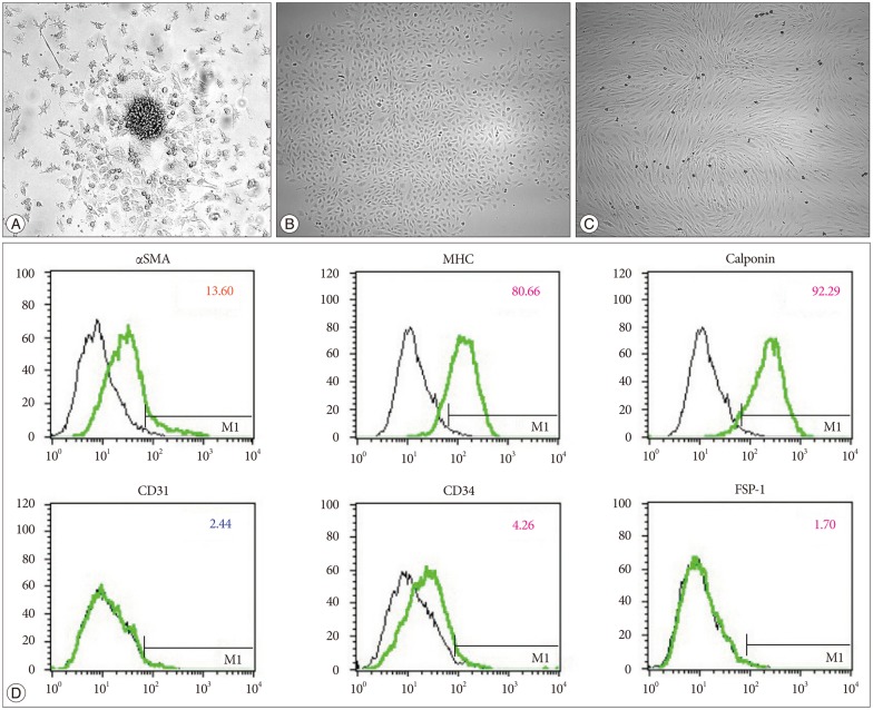
Fig. 1 Vascular progenitor cells. Values on the y axis quantify the intensity of fluorescein isothiocyanate staining (FITC log), and on the x axis the values represent the number of cells counted. A : Early endothelial progenitor cells (EPCs), also called colony-forming units or cell clusters (×200). This cell cluster is composed of a central core of round cells that are surrounded by spindle shaped cells. B : Late EPCs, also known as endothelial outgrowth cells (×40). The cells are arranged in cobblestone-like formations. C : Smooth muscle progenitor cells (SPCs) (×40). These cells appear elon-gated and have a typical hill-and-valley appearance. D : Fluorescence-activated cell sorter analysis of the cells shown in C. The cells express smooth muscle myosin heavy chain (MHC) and calponin, which are smooth-muscle specific, with little expression of CD31 and fibroblast-specific protein 1 (FSP-1). αSMA : smooth muscle actin-alpha.
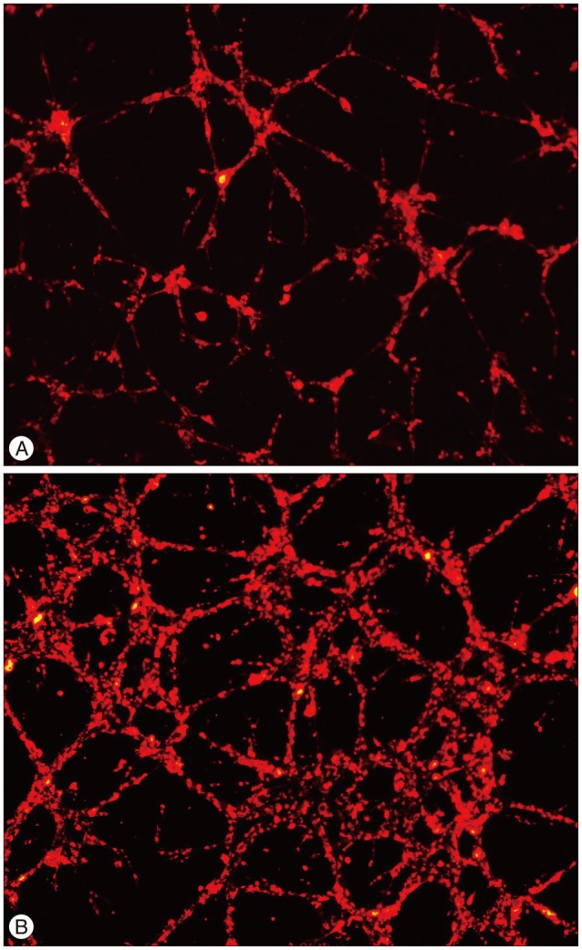
Fig. 2 Vascular progenitor cells. Values on the y axis quantify the intensity of fluorescein isothiocyanate staining (FITC log), and on the x axis the values represent the number of cells counted. A : Early endothelial progenitor cells (EPCs), also called colony-forming units or cell clusters (×200). This cell cluster is composed of a central core of round cells that are surrounded by spindle shaped cells. B : Late EPCs, also known as endothelial outgrowth cells (×40). The cells are arranged in cobblestone-like formations. C : Smooth muscle progenitor cells (SPCs) (×40). These cells appear elon-gated and have a typical hill-and-valley appearance. D : Fluorescence-activated cell sorter analysis of the cells shown in C. The cells express smooth muscle myosin heavy chain (MHC) and calponin, which are smooth-muscle specific, with little expression of CD31 and fibroblast-specific protein 1 (FSP-1). αSMA : smooth muscle actin-alpha.
Cited by 2 articles
-
A Recent Update of Clinical and Research Topics Concerning Adult Moyamoya Disease
Jin Pyeong Jeon, Jeong Eun Kim
J Korean Neurosurg Soc. 2016;59(6):537-543. doi: 10.3340/jkns.2016.59.6.537.Cyclin-Dependent Kinase Inhibitor 2A is a Key Regulator of Cell Cycle Arrest and Senescence in Endothelial Colony-Forming Cells in Moyamoya Disease
Seung Ah Choi, Youn Joo Moon, Eun Jung Koh, Ji Hoon Phi, Ji Yeoun Lee, Kyung Hyun Kim, Seung-Ki Kim
J Korean Neurosurg Soc. 2023;66(6):642-651. doi: 10.3340/jkns.2023.0005.
Reference
-
1. Asahara T, Murohara T, Sullivan A, Silver M, van der Zee R, Li T, et al. Isolation of putative progenitor endothelial cells for angiogenesis. Science. 1997; 275:964–967. PMID: 9020076.
Article2. Chu K, Jung KH, Lee ST, Park HK, Sinn DI, Kim JM, et al. Circulating endothelial progenitor cells as a new marker of endothelial dysfunction or repair in acute stroke. Stroke. 2008; 39:1441–1447. PMID: 18356550.
Article3. Fadini GP, Losordo D, Dimmeler S. Critical reevaluation of endothelial progenitor cell phenotypes for therapeutic and diagnostic use. Circ Res. 2012; 110:624–637. PMID: 22343557.
Article4. Gill M, Dias S, Hattori K, Rivera ML, Hicklin D, Witte L, et al. Vascular trauma induces rapid but transient mobilization of VEGFR2(+)AC133(+) endothelial precursor cells. Circ Res. 2001; 88:167–174. PMID: 11157668.
Article5. Jung KH, Chu K, Lee ST, Park HK, Kim DH, Kim JH, et al. Circulating endothelial progenitor cells as a pathogenetic marker of moyamoya disease. J Cereb Blood Flow Metab. 2008; 28:1795–1803. PMID: 18612318.
Article6. Kang HS, Moon YJ, Kim YY, Park WY, Park AK, Wang KC, et al. Smooth-muscle progenitor cells isolated from patients with moyamoya disease : novel experimental cell model. J Neurosurg. 2014; 120:415–425. PMID: 24160477.
Article7. Khakoo AY, Finkel T. Endothelial progenitor cells. Annu Rev Med. 2005; 56:79–101. PMID: 15660503.
Article8. Kim JH, Jung JH, Phi JH, Kang HS, Kim JE, Chae JH, et al. Decreased level and defective function of circulating endothelial progenitor cells in children with moyamoya disease. J Neurosci Res. 2010; 88:510–518. PMID: 19774676.
Article9. Kim SK, Cho BK, Phi JH, Lee JY, Chae JH, Kim KJ, et al. Pediatric moyamoya disease : an analysis of 410 consecutive cases. Ann Neurol. 2010; 68:92–101. PMID: 20582955.10. Rafat N, Beck GCh, Peña-Tapia PG, Schmiedek P, Vajkoczy P. Increased levels of circulating endothelial progenitor cells in patients with Moyamoya disease. Stroke. 2009; 40:432–438. PMID: 19095988.
Article11. Shintani S, Murohara T, Ikeda H, Ueno T, Honma T, Katoh A, et al. Mobilization of endothelial progenitor cells in patients with acute myocardial infarction. Circulation. 2001; 103:2776–2779. PMID: 11401930.
Article12. Simper D, Stalboerger PG, Panetta CJ, Wang S, Caplice NM. Smooth muscle progenitor cells in human blood. Circulation. 2002; 106:1199–1204. PMID: 12208793.
Article
- Full Text Links
-
- Actions
-
Cited
- CITED
-
- Close
- Share
-
- Similar articles
-
- The Pathophysiology of Moyamoya Disease: An Update
- Adult-Onset Moyamoya Disease Manifesting as Vascular Parkinsonism
- Circulating Endothelial Progenitor Cells and Vasculogenic Factors in Pterygium Pathogenesis
- Neuroimaging Diagnosis and Treatment of Moyamoya Disease
- Moyamoya-like Disease Associated with Intracranial Aneurysm
