A Case of Ectopic Rathke's Cleft Cyst in the Prepontine Cistern
- Affiliations
-
- 1Department of Neurosurgery, Keimyung University School of Medicine, Dongsan Medical Center, Daegu, Korea. bach1158@dsmc.or.kr
- KMID: 2190556
- DOI: http://doi.org/10.3340/jkns.2012.52.2.152
Abstract
- A Rathke's cleft cyst (RCC) is a benign pituitary cyst derived from the remnant of Rathke's pouch, and usually presents as an intrasellar lesion with varying degrees of suprasellar extension. However, to date, a description of a primary prepontine RCC with no intrasellar component has not been reported. The author describes an exceptional case of a symptomatic RCC located behind the sella turcica in a 41-year-old woman who presented with severe headache. The author also provides an embryological hypothesis of the development of an ectopic RCC, with a special emphasis on radiologic characteristics.
Figure
-
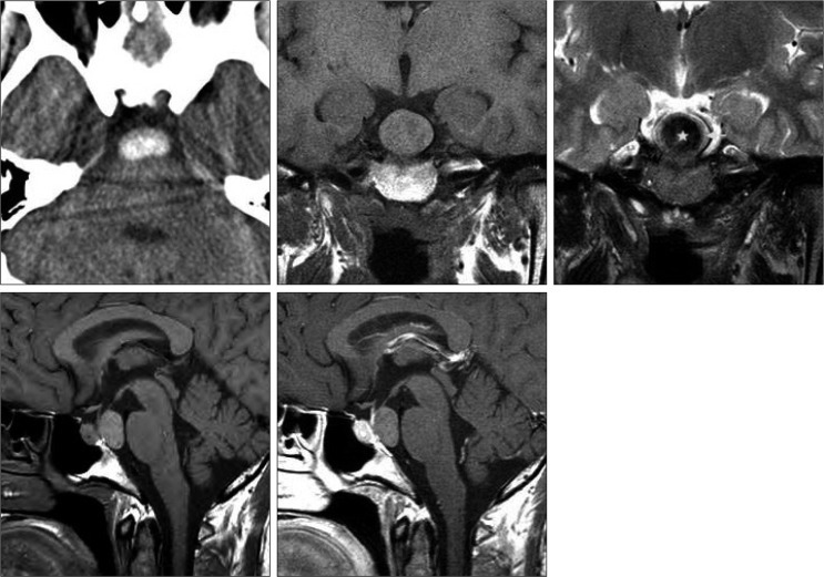
Fig. 1 Neuroimaging studies of an ectopic Rathke's cleft cyst. Computed tomography scans show a homogenous oval-shaped high density in the prepontine cistern. Magnetic resonance images depict a nonenhancing retrosellar mass with an intracystic nodule that displays iso- to slight hyperintensity on T1-weighted sequence and hypointensity on T2-weighted sequence. The nodule (*) is surrounded by thin isointense cyst fluid (arrow).
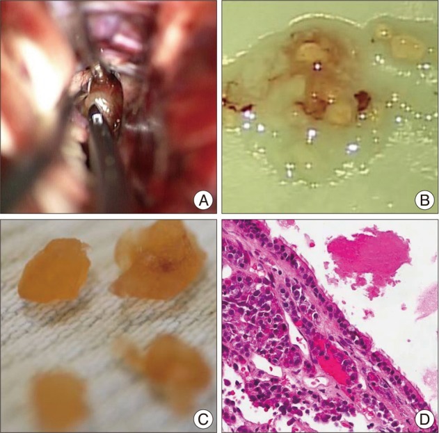
Fig. 2 Operative and pathology features of a Rathke's cleft cyst in the prepontine cistern. Intraoperative view demonstrates resection of a cyst wall with thick mucoid and white-yellow substances (A). Photomicrographs of surgical specimens show the cystic contents and the waxy nodules (B and C). Histopathological examination of cyst wall illustrates ciliated columnar epitheliums and inflammatory infiltrates (H&E, original magnification ×100) (D).

Fig. 3 Postoperative computed tomography scans exhibit disappearance of the hyperdense cyst behind the pituitary fossa.
Reference
-
1. Aho CJ, Liu C, Zelman V, Couldwell WT, Weiss MH. Surgical outcomes in 118 patients with Rathke cleft cysts. J Neurosurg. 2005; 102:189–193. PMID: 15739543.
Article2. Anand VK, Osborne CM, Harkey HL 3rd. Infiltrative clival pituitary adenoma of ectopic origin. Otolaryngol Head Neck Surg. 1993; 108:178–183. PMID: 8441545.
Article3. Barrow DL, Spector RH, Takei Y, Tindall GT. Symptomatic Rathke's cleft cysts located entirely in the suprasellar region : review of diagnosis, management, and pathogenesis. Neurosurgery. 1985; 16:766–772. PMID: 4010898.
Article4. Binning MJ, Gottfried ON, Osborn AG, Couldwell WT. Rathke cleft cyst intracystic nodule : a characteristic magnetic resonance imaging finding. J Neurosurg. 2005; 103:837–840. PMID: 16304987.
Article5. Chen CJ. Suprasellar and infrasellar craniopharyngioma with a persistent craniopharyngeal canal: case report and review of the literature. Neuroradiology. 2001; 43:760–762. PMID: 11594427.
Article6. Choi SH, Kwon BJ, Na DG, Kim JH, Han MH, Chang KH. Pituitary adenoma, craniopharyngioma, and Rathke cleft cyst involving both intrasellar and suprasellar regions : differentiation using MRI. Clin Radiol. 2007; 62:453–462. PMID: 17398271.
Article7. Chuang CC, Chen YL, Jung SM, Pai PC. A giant retroclival Rathke's cleft cyst. J Clin Neurosci. 2010; 17:1189–1191. PMID: 20627584.
Article8. Hanna E, Weissman J, Janecka IP. Sphenoclival Rathke's cleft cysts : embryology, clinical appearance and management. Ear Nose Throat J. 1998; 77:396–399. PMID: 9615520.
Article9. Kim JE, Kim JH, Kim OL, Paek SH, Kim DG, Chi JG, et al. Surgical treatment of symptomatic Rathke cleft cysts : clinical features and results with special attention to recurrence. J Neurosurg. 2004; 100:33–40. PMID: 14743909.
Article10. Kleinschmidt-DeMasters BK, Lillehei KO, Stears JC. The pathologic, surgical, and MR spectrum of Rathke cleft cysts. Surg Neurol. 1995; 44:19–26. discussion 26-27. PMID: 7482247.
Article11. Megdiche-Bazarbacha H, Ben Hammouda K, Aicha AB, Sebai R, Belghith L, Khaldi M, et al. Intrasphenoidal rathke cleft cyst. AJNR Am J Neuroradiol. 2006; 27:1098–1100. PMID: 16687551.12. Nishioka H, Haraoka J, Izawa H, Ikeda Y. Magnetic resonance imaging, clinical manifestations, and management of Rathke's cleft cyst. Clin Endocrinol (Oxf). 2006; 64:184–188. PMID: 16430718.
Article13. Osborn AG, Preece MT. Intracranial cysts : radiologic-pathologic correlation and imaging approach. Radiology. 2006; 239:650–664. PMID: 16714456.
Article14. Panagopoulos KP, Jolesz FA, el-Azouzi M, Black PM. Mucinous cysts of the pituitary stalk. Report of two cases. J Neurosurg. 1989; 71:276–278. PMID: 2746351.15. Ross DA, Norman D, Wilson CB. Radiologic characteristics and results of surgical management of Rathke's cysts in 43 patients. Neurosurgery. 1992; 30:173–178. discussion 178-179. PMID: 1545884.
Article
- Full Text Links
-
- Actions
-
Cited
- CITED
-
- Close
- Share
-
- Similar articles
-
- Symptomatic Rathke's Cleft Cyst in the Interpeduncular Cistern: Case Report
- Suprasellar Rathke Cleft Cyst: A case report
- Rathke's Cleft Cyst: Case Report
- Large Ossified Rathke's Cleft Cyst: A Case Report and Review of the Literature
- Pituitary Tumors Composed of Adenohypophysial Adenoma and Rathke's Cleft Cyst Elements
