Clinical and Radiological Findings of Nerve Root Herniation after Discectomy of Lumbar Disc Herniation
- Affiliations
-
- 1Department of Neurosurgery, Wooridul Spine Hospital, Seoul, Korea. spinejjs@yahoo.co.kr
- 2Department of Neurosurgery, Seoul Wooridul Hospital, Seoul, Korea.
- KMID: 2190527
- DOI: http://doi.org/10.3340/jkns.2012.52.1.62
Abstract
- The authors report 2 cases of nerve root herniation after discectomy of a large lumbar disc herniation caused by an unrecognized dural tear. Patients complained of the abrupt onset of radiating pain after lumbar discectomy. Magnetic resonance imaging showed cerebrospinal fluid signal in the disc space and nerve root displacement into the disc space. Symptoms improved after the herniated nerve root was repositioned. Clinical symptoms and suggestive radiologic image findings are important for early diagnosis and treatment.
Keyword
Figure
-
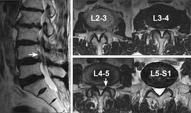
Fig. 1 Preoperative magnetic resonance image showing very large, upwardly migrated disc herniation (arrow) at the left L4-5 level; spinal stenosis at the L2-3, L3-4, and L4-5 levels; and mild lateral recess stenosis at the left L5-S1 level.
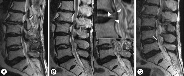
Fig. 2 A : After discectomy at the L4-5 level, magnetic resonance image (MRI) shows complete removal of the herniated disc. Note the showed cerebrospinal fluid signal in the L4-5 disc space, which is suggestive of dural tear. B : MRI taken when the patient complained of newly developed sciatic pain after discharge. Left sagittal MRI shows no significant interval change from the immediate postoperative MRI (A). However, a nerve root had herniated (arrowheads) into the intervertebral disc space, as shown by the gull wing shape. C : MRI after additional decompression at the left L3-4 and L5-S1 levels. Note the gull wing-shaped nerve root herniation (arrow), which contacts the intervertebral disc.

Fig. 3 MRI showing nerve root herniation (arrows) into the disc space. The point where the nerve root meets with the intervertebral disc space shows a darker signal than that in Fig. 2C. The nerve root formed a loop inside the annulus (arrowheads) on the T2-weighted axial scan.
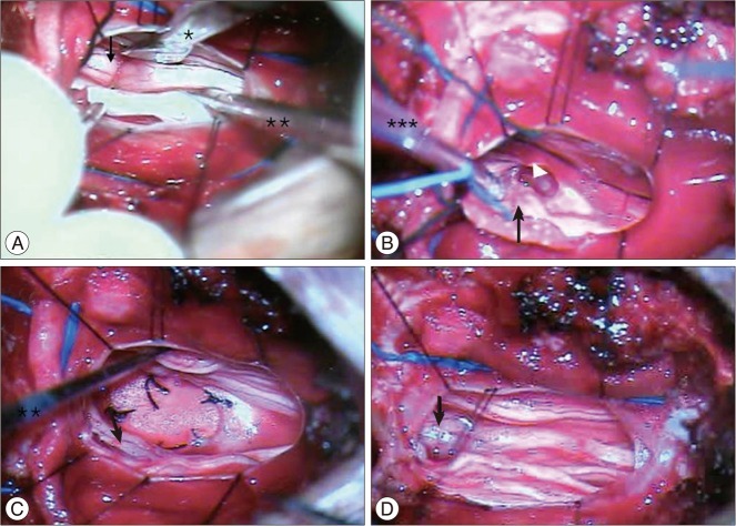
Fig. 4 A : Intraoperative microscopic photo (right : cranial, left : caudal, upper : left, lower : right) showing the nerve root herniated (arrow) into the disc space (*root retractor, **microprobe). B : Severe adhesion (arrowhead) is observed between the nerve root, dura, and disc (*** suction). C : After release of the nerve root, the ventral dural defect is closed with artificial dura. D : The nerve root was repositioned after dural repair. Note the swelling of the nerve root due to ample manipulation for adhesiolysis.

Fig. 5 Preoperative magnetic resonance imaging showing paramedian extruded disc herniation (arrow) at the left L2-3 level and spinal stenosis at the L3-4 and L4-5 levels.
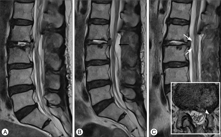
Fig. 6 Magnetic resonance image performed after additional decompression at the L3-4 and L4-5 levels. The attachment of the nerve root at the L2-3 disc space (black arrowhead) and showed cerebrospinal fluid signal in the L2-3 disc space, which are suggestive of dural tear, can be seen. B : Follow-up MRI showing an epidural hematoma at the L2-3 level (black arrow), which was removed by computed tomography-guided needle aspiration. C : MRI after aspiration of the epidural hematoma. The attachment of the ventral nerve root at the L2-3 disc space is seen in the shape of a gull wing (white arrows) on the sagittal scan, and the attachment of the rootlets on the ventral dura can be seen on the axial scan (white arrowhead).
Reference
-
1. Ahn Y, Lee HY, Lee SH, Lee JH. Dural tears in percutaneous endoscopic lumbar discectomy. Eur Spine J. 2011; 20:58–64. PMID: 20582555.
Article2. Bosacco SJ, Gardner MJ, Guille JT. Evaluation and treatment of dural tears in lumbar spine surgery : a review. Clin Orthop Relat Res. 2001; 238–247. PMID: 11501817.3. Cammisa FP Jr, Girardi FP, Sangani PK, Parvataneni HK, Cadag S, Sandhu HS. Incidental durotomy in spine surgery. Spine (Phila Pa 1976). 2000; 25:2663–2667. PMID: 11034653.
Article4. Hodges SD, Humphreys SC, Eck JC, Covington LA. Management of incidental durotomy without mandatory bed rest. A retrospective review of 20 cases. Spine (Phila Pa 1976). 1999; 24:2062–2064. PMID: 10528385.5. Jones AA, Stambough JL, Balderston RA, Rothman RH, Booth RE Jr. Long-term results of lumbar spine surgery complicated by unintended incidental durotomy. Spine (Phila Pa 1976). 1989; 14:443–446. PMID: 2718049.
Article6. Nishi S, Hashimoto N, Takagi Y, Tsukahara T. Herniation and entrapment of a nerve root secondary to an unrepaired small dural laceration at lumbar hemilaminectomies. Spine (Phila Pa 1976). 1995; 20:2576–2579. PMID: 8610254.
Article7. Pavlou G, Bucur SD, van Hille PT. Entrapped spinal nerve roots in a pseudomeningocoele as a complication of previous spinal surgery. Acta Neurochir (Wien). 2006; 148:215–219. discussion 219-220. PMID: 16374564.
Article8. Saxler G, Krämer J, Barden B, Kurt A, Pförtner J, Bernsmann K. The long-term clinical sequelae of incidental durotomy in lumbar disc surgery. Spine (Phila Pa 1976). 2005; 30:2298–2302. PMID: 16227893.
Article9. Sin AH, Caldito G, Smith D, Rashidi M, Willis B, Nanda A. Predictive factors for dural tear and cerebrospinal fluid leakage in patients undergoing lumbar surgery. J Neurosurg Spine. 2006; 5:224–227. PMID: 16961083.
Article10. Töppich HG, Feldmann H, Sandvoss G, Meyer F. Intervertebral space nerve root entrapment after lumbar disc surgery. Two cases. Spine (Phila Pa 1976). 1994; 19:249–250. PMID: 8153836.
- Full Text Links
-
- Actions
-
Cited
- CITED
-
- Close
- Share
-
- Similar articles
-
- Conjoined Nerve Root Anomaly Associated with Lumbar Disc Herniation: A Case Report
- A Case of Nerve-Root Anomaly Associated with Herniated Intervertebral Disc in Lumbar: Case Report
- Redundant Nerve Roots of Cauda Equina Mimicking Intradural Disc Herniation: A Case Report
- Spinal Nerve Root Swelling Mimicking Intervertebral Disc Herniation in Magnetic Resonance Imaging: A Case Report
- Minimally Invasive Surgery in Lumbar Disc Herniations with MicroEndoscopic Discectomy System: Technical Note
