Midline-Splitting Open Door Laminoplasty Using Hydroxyapatite Spacers : Comparison between Two Different Shaped Spacers
- Affiliations
-
- 1Department of Neurological Surgery, Gangneung Asan Medical Center, University of Ulsan College of Medicine, Gangneung, Korea.
- 2Department of Neurological Surgery, Asan Medical Center, University of Ulsan College of Medicine, Seoul, Korea. srjeon@amc.seoul.kr
- KMID: 2190519
- DOI: http://doi.org/10.3340/jkns.2012.52.1.27
Abstract
OBJECTIVE
Although hydroxyapatite (HA) spacer has been used for laminoplasty, there have been no reports on factors associated with fusion and on the effects of HA shape.
METHODS
During January 2004 and January 2010, 45 patients with compressive cervical myelopathy underwent midline-splitting open door laminoplasty with winged (33 cases) and wingless (12 cases) HAs by a single surgeon. Minimal and mean follow up times were 12 and 28.1 months, respectively. Japanese Orthopedic Association (JOA) score was used for clinical outcome measurement. Cervical X-rays were taken preoperatively, immediately post-operatively, and after 3, 6, and 12 months and computed tomography scans were performed preoperatively, immediately post-operatively and after 12 months. Cervical lordosis, canal dimension, fusion between lamina and HA, and affecting factors of fusion were analyzed.
RESULTS
All surgeries were performed on 142 levels, 99 in the winged and 43 in the wingless HA groups. JOA scores of the winged group changed from 10.4+/-2.94 to 13.3+/-2.35 and scores of the wingless group changed from 10.8+/-2.87 to 13.8+/-3.05. There was no significant difference on lordotic and canal dimensional change between two groups. Post-operative 12 month fusion rate between lamina and HA was significantly lower in the winged group (18.2 vs. 48.8% p=0.001). Multivariate analysis showed that ossification of the posterior longitudinal ligament, male gender, and wingless type HA were significantly associated with fusion.
CONCLUSION
Clinical outcome was similar in patients receiving winged and wingless HA, but the wingless type was associated with a higher rate of fusion between HA and lamina at 12 months post-operatively.
MeSH Terms
Figure
-
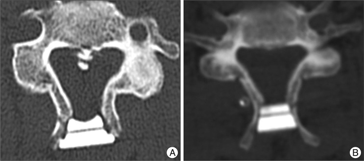
Fig. 1 Types of HA. A : Winged type. B : Wingless type. HA : hydroxyapatite.
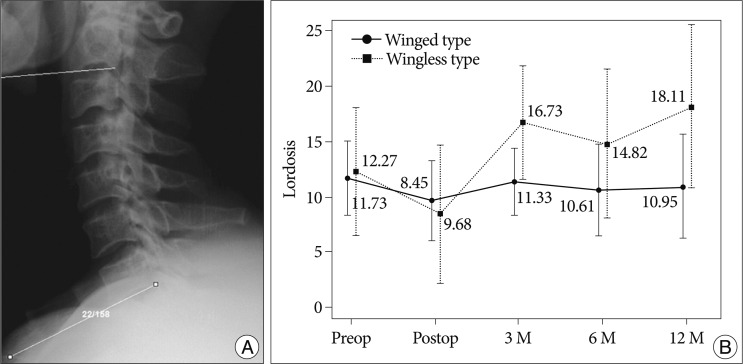
Fig. 2 A : Cervical lordosis, defined as the angle between C2 and C7 inferior end plates. B : Changes in cervical lordosis in the winged and wingless groups. Lordosis changed from 11.73 to 10.95 degrees in the winged type group and from 12.27 to 18.11 degrees in the wingless type group (p=0.135).
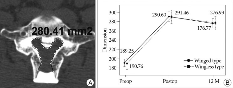
Fig. 3 A : Spinal canal dimension measurements. B : The spinal canal dimensions increased immediately post-operatively in both groups, from 190.76 to 291.46 in the winged group and from 189.25 to 290.60 in the winged group, but decreased to 276.77 and 276.93, respectively, after 12 months (p=0.970).
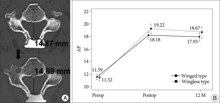
Fig. 4 A : AP distance, as measured on CT axial image. (Upper image) immediate post-operative AP distance; (lower image) AP distance after 12 months. B : The AP distance of the spinal canal increased immediately after surgery from 11.52 mm to 19.22 mm in the wingless group and from 11.59 mm to 18.18 mm in the winged type group. After 12 months, the AP distances were 18.67 mm and 17.93 mm in the wingless and winged groups, respectively (p=0.021). AP : anterior-posterior, CT : computed tomography.
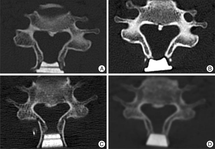
Fig. 5 A : Immediate post-operative axial CT image in the winged type group showing good touch status. B : Post-operative 12 month axial CT in the same patient showing non-fusion and spinous process resorption. C : Immediate post-operative axial CT image in the wingless type group showing good touch status. D : Post-operative 12 month axial CT in the same patient showing fusion, but spinous process resorption. CT : computed tomography.
Cited by 1 articles
-
A Comparison of Implants Used in Double Door Laminoplasty : Allogeneic Bone Spacer versus Hydroxyapatite Spacer
Dong Yoon Lee, Chang Kyu Lee, In-Soo Kim
J Korean Neurosurg Soc. 2016;59(6):604-609. doi: 10.3340/jkns.2016.59.6.604.
Reference
-
1. Chiba K, Ogawa Y, Ishii K, Takaishi H, Nakamura M, Maruiwa H, et al. Long-term results of expansive open-door laminoplasty for cervical myelopathy--average 14-year follow-up study. Spine (Phila Pa 1976). 2006; 31:2998–3005. PMID: 17172996.
Article2. Hirabayashi S, Kumano K. Contact of hydroxyapatite spacers with split spinous processes in double-door laminoplasty for cervical myelopathy. J Orthop Sci. 1999; 4:264–268. PMID: 10436273.
Article3. Hoshi K, Kurokawa T, Nakamura K, Hoshino Y, Saita K, Miyoshi K. Expansive cervical laminoplasties--observations on comparative changes in spinous process lengths following longitudinal laminal divisions using autogenous bone or hydroxyapatite spacers. Spinal Cord. 1996; 34:725–728. PMID: 8961430.
Article4. Iguchi T, Kanemura A, Kurihara A, Kasahara K, Yoshiya S, Doita M, et al. Cervical laminoplasty : evaluation of bone bonding of a high porosity hydroxyapatite spacer. J Neurosurg. 2003; 98:137–142. PMID: 12650397.
Article5. Iwasaki M, Kawaguchi Y, Kimura T, Yonenobu K. Long-term results of expansive laminoplasty for ossification of the posterior longitudinal ligament of the cervical spine : more than 10 years follow up. J Neurosurg. 2002; 96:180–189. PMID: 12450281.
Article6. Kaito T, Hosono N, Makino T, Kaneko N, Namekata M, Fuji T. Postoperative displacement of hydroxyapatite spacers implanted during double-door laminoplasty. J Neurosurg Spine. 2009; 10:551–556. PMID: 19558287.
Article7. Kawaguchi Y, Kanamori M, Ishihara H, Ohmori K, Nakamura H, Kimura T. Minimum 10-year followup after en bloc cervical laminoplasty. Clin Orthop Relat Res. 2003; 129–139. PMID: 12782868.
Article8. Kimura A, Seichi A, Inoue H, Hoshino Y. Long-term results of double-door laminoplasty using hydroxyapatite spacers in patients with compressive cervical myelopathy. Eur Spine J. 2011; 20:1560–1566. PMID: 21336508.
Article9. Kimura I, Shingu H, Nasu Y. Long-term follow-up of cervical spondylotic myelopathy treated by canal-expansive laminoplasty. J Bone Joint Surg Br. 1995; 77:956–961. PMID: 7593114.
Article10. Kokubun S, Kashimoto O, Tanaka Y. Histological verification of bone bonding and ingrowth into porous hydroxyapatite spinous process spacer for cervical laminoplasty. Tohoku J Exp Med. 1994; 173:337–344. PMID: 7846685.
Article11. Kong Q, Zhang L, Liu L, Li T, Gong Q, Zeng J, et al. Effect of the decompressive extent on the magnitude of the spinal cord shift after expansive open-door laminoplasty. Spine (Phila Pa 1976). 2011; 36:1030–1036. PMID: 21150700.
Article12. Kubo S, Goel VK, Yang SJ, Tajima N. Biomechanical evaluation of cervical double-door laminoplasty using hydroxyapatite spacer. Spine (Phila Pa 1976). 2003; 28:227–234. PMID: 12567022.
Article13. Kurokawa T, Tsuyama N, Tanaka H, Kobayashi M, Machida H, Nakamura K, et al. [Double-open door laminoplasty]. Bessatsu Seikeigeka. 1982; 2:234–240.14. Martin-Benlloch JA, Maruenda-Paulino JI, Barra-Pla A, Laguia-Garzaran M. Expansive laminoplasty as a method for managing cervical multilevel spondylotic myelopathy. Spine (Phila Pa 1976). 2003; 28:680–684. PMID: 12671355.
Article15. Nakano K, Harata S, Suetsuna F, Araki T, Itoh J. Spinous process-splitting laminoplasty using hydroxyapatite spinous process spacer. Spine (Phila Pa 1976). 1992; 17:S41–S43. PMID: 1314432.
Article16. Ogawa Y, Chiba K, Matsumoto M, Nakamura M, Takaishi H, Hirabayashi H, et al. a comparison with nonsegmental-type lesions. J Neurosurg Spine. 2005; 3:198–204. PMID: 16235702.17. Ratliff JK, Cooper PR. Cervical laminoplasty : a critical review. J Neurosurg. 2003; 98:230–238. PMID: 12691377.18. Sakaura H, Hosono N, Mukai Y, Ishii T, Iwasaki M, Yoshikawa H. Long-term outcome of laminoplasty for cervical myelopathy due to disc herniation : a comparative study of laminoplasty and anterior spinal fusion. Spine (Phila Pa 1976). 2005; 30:756–759. PMID: 15803077.
Article
- Full Text Links
-
- Actions
-
Cited
- CITED
-
- Close
- Share
-
- Similar articles
-
- Novel Hybrid Hydroxyapatite Spacers Ensure Sufficient Bone Bonding in Cervical Laminoplasty
- Comparison of Early Surgical Outcome between Unilateral Open-Door Laminoplasty and Midline Splitting Laminoplasty
- Modified Open-door Laminoplasty Using Hydroxyapatite Spacers and Miniplates
- Midline Splitting Cervical Laminoplasty Using Allogeneic Bone Spacers: Comparison of Fusion Rates between Cervical Spondylotic Myelopathy and Ossification of Posterior Longitudinal Ligament
- Is the Cross-sectional Area after Unilateral Open Door Laminoplasty Wider than that after Midline Splitting Laminoplasty ? : Mathematical Approach
